Authors: Andrew Klein & Sera Elizabeth Klein
Dedication: To every patient who has suffered through a failed implant. To every family that has watched a loved one undergo repeat surgeries. To every surgeon who has had to tell a patient that the device they trusted has failed. And to the engineers, scientists, and dreamers who believe we can build a better way.
Abstract
This paper examines the systemic failures of current heart valve prostheses—mechanical, bioprosthetic, and transcatheter—and proposes a new paradigm: open-source, patient-specific, bioresorbable heart valves. We document the clinical and economic costs of existing devices: mechanical valves that require lifelong anticoagulation with significant bleeding and thromboembolic risks; bioprosthetic valves that degenerate within 5–15 years, often requiring repeat surgery; and transcatheter valves that show concerning rates of hemodynamic deterioration. We then present the emerging technologies that make a better alternative possible: the ParaValve open-source framework for parametric design and fluid-structure interaction simulation; 3D-printed bioresorbable materials that promote tissue regeneration; and patient-specific geometry enabled by additive manufacturing. We propose a design framework that integrates these technologies to create heart valves that are durable, biocompatible, and capable of being regenerated by the patient’s own body—rendering the current extractive model of medical device manufacturing obsolete.
1. Introduction: The Failure of the Current System
Heart valve disease affects millions worldwide. In the United States alone, more than 5 million people are diagnosed with heart valve disease annually. The standard of care—valve replacement—offers no effective long-term solution. Most replacement valves are made of animal tissue and last only 10 to 15 years before they must be replaced. For pediatric patients, who grow and change, the situation is even more dire: solutions are extremely limited and can require multiple reinterventions.
The current system is extractive. Patients pay—with their health, their finances, and their lives—for devices that are rushed to market, inadequately tested, and designed for profit rather than durability. The same pattern we have documented in other medical devices—aggressive marketing, minimal oversight, externalised costs—is replicated in the heart valve industry.
This paper proposes an alternative: open-source, patient-specific, bioresorbable heart valves designed using parametric modeling, 3D printing, and tissue engineering. This is not a marginal improvement. It is a paradigm shift.
2. The Problems with Mechanical Valves
Mechanical heart valves (MHVs) are made from rigid materials such as pyrolytic carbon. They offer excellent durability but require lifelong anticoagulation due to thromboembolic risk.
2.1 The Anticoagulation Burden
For patients with a mechanical aortic valve, warfarin is the only approved drug for anticoagulation. Patients face numerous challenges inherent in its use—including multiple food and drug interactions and the need for frequent blood monitoring. Many patients, especially younger ones, opt for a biological valve instead—despite knowing it will be less durable.
2.2 The PROACT Xa Trial Failure
The PROACT Xa trial was designed to determine if apixaban—a factor Xa inhibitor—could be a suitable alternative to warfarin for patients with an On-X mechanical aortic valve. The trial was halted prematurely in September 2022 due to an excess risk of blood clots.
The results were devastating:
· 20 valve thrombosis or thromboembolism events in the apixaban group (4.2%/patient-year) compared to 6 in the warfarin group
· 14 thromboembolic strokes occurred with apixaban compared to none with warfarin
As Dr Lars Svensson, co-chair of the PROACT Xa steering committee, concluded: “We had good reason to believe that apixaban could be a suitable alternative to warfarin… Unfortunately, the data did not support this”.
2.3 The Clinical Reality
A 59-year-old woman with a mechanical valve suffered recurrent embolic strokes nearly 7 years after implantation. The PROACT Xa trial results raised questions about the concomitant use of aspirin, which did not reduce thromboembolism risk but increased major bleeding.
Mechanical valves trade durability for a lifetime of anticoagulation—and even then, the risk of stroke and bleeding remains.
3. The Problems with Bioprosthetic Valves
Bioprosthetic heart valves (BHVs) offer improved hemodynamics and lower thrombogenicity but suffer from structural degeneration and limited durability, particularly in younger patients.
3.1 The Trifecta Valve Failure
The Trifecta family of bioprosthetic aortic valves—manufactured by Abbott—has been the subject of serious warnings. In February 2023, both Abbott and the FDA warned that the Trifecta valves may begin deteriorating within the first five years of implantation, potentially requiring earlier-than-expected replacement or even leading to death.
Real-world data told a different story from clinical trials:
· Clinical trials showed peak structural valve deterioration at the eight-year mark
· Real-world complaints showed peak deterioration at the three- to four-year mark
· The majority of early deterioration cases centred on a tear of at least one leaflet
· Later failures were caused by calcification and hardening
3.2 Early Structural Valve Degeneration
A study reported 3 cases of early SVD after aortic valve replacement with the Trifecta GT, for a cumulative incidence of 3.3% at 3.5 years. The SVD occurred between 1.9 and 3.2 years after implantation, with all patients presenting with aortic insufficiency and heart failure. Intraoperative findings revealed a detached prosthetic leaflet at the stent post.
The authors postulated that the externally mounted leaflet design and mechanical abrasion from the aortic wall may have contributed to the early failure. A greater degree of prosthetic leaflet flutter was observed.
3.3 The Trifecta GT: Innovation Is Not Always Progress
As one commentary aptly titled: “Innovation is not always progress“. The newer-generation Trifecta GT was designed to improve results using novel anti-leaflet calcification technology—yet it failed. Structural valve degeneration remains a major disadvantage of bioprosthetic aortic valve replacement.
Bioprosthetic valves trade short-term comfort for long-term failure—and patients pay the price with repeat surgeries.
4. The Problems with TAVR Valves
Transcatheter aortic valve replacement (TAVR) has become an established therapy for severe aortic stenosis, with valve durability demonstrated for at least five years (and up to eight years) in recent studies. However, concerns remain.
4.1 Hemodynamic Valve Deterioration
Unlike mechanical valves, bioprosthetic valves used in TAVR are susceptible to hemodynamic valve deterioration (HVD) over time, driven by structural changes such as leaflet thickening, calcification, and tearing.
The incidence of moderate or severe HVD:
· 2.2% at 1 year
· 10.8% at 5 years
· 25.6% at 10 years
Severe HVD occurred in 0.5%, 2.6%, and 12.7% of patients at the respective time points. Patients with HVD had a 5-fold increased risk for requiring aortic valve reintervention within 5 years.
4.2 Early Structural Deterioration
While structural valve deterioration within 5 years is rare, it does occur. An 88-year-old man developed progressive dyspnoea and chest pain five years after TAVR, with examination revealing severe aortic regurgitation. He required surgical aortic valve replacement.
This case underscores the possibility of rapid prosthetic valve failure and highlights the ongoing importance of physical examination in detecting severe regurgitation.
4.3 Native Leaflet Calcification as a Predictor
Independent predictors of HVD included aortic valve calcium volume and residual aortic regurgitation at discharge, marking the degree of native aortic valve calcification as a novel predictor of TAVR valve degeneration. Heavily calcified native aortic valves can hinder optimal device expansion, leading to an underexpanded prosthesis that increases leaflet stress and accelerates structural deterioration.
TAVR valves offer less invasive surgery—but at the cost of uncertain long-term durability.
5. The Promise of Open-Source Design
5.1 The ParaValve Framework
ParaValve is an open-source framework for parametric design and fluid–structure interaction simulation of bioprosthetic heart valves in patient-specific aortic geometries.
Key features:
· NURBS-based procedural modelling enables patient-specific aortic geometry creation
· Efficient fluid–structure interaction framework for bioprosthetic valve analysis
· Enables rapid, personalised design iterations of bioprosthetic heart valves
· Open-source modelling framework for patient-specific cardiac flow simulations
By enabling a modular and expandable workflow, the framework supports iterative optimisation of valve designs to achieve improved haemodynamic performance and durability. It offers researchers and engineers a streamlined pathway toward innovative and patient-specific cardiovascular solutions.
5.2 Why Open-Source Matters
The current medical device industry is built on secrecy. Designs are proprietary. Testing data is hidden. Failures are buried. Patients are left with no way to know whether the device they are receiving has been adequately tested—or whether it is yet another failure waiting to happen.
Open-source design changes this:
· Designs are transparent and auditable
· Testing data is public and verifiable
· Failures are learned from, not hidden
· Innovation is accelerated by collaboration
· Costs are reduced by eliminating proprietary markups
6. Biomaterials for the Next Generation
6.1 Bioresorbable Polymers: A Paradigm Shift
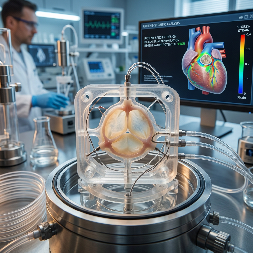
Georgia Tech researchers have created a 3D-printed heart valve made of bioresorbable materials designed to fit an individual patient’s unique anatomy. Once implanted, the valves are absorbed by the body and replaced by new tissue that performs the function the device once served.
The material: The valve is 3D-printed using a biocompatible material called poly(glycerol dodecanedioate). It has shape memory, so it can be folded and delivered via a catheter rather than open heart surgery. Once it is implanted and reaches body temperature, the device refolds into its original shape. The material then signals to the body to make its own new tissue.
Why this matters:
· Current animal tissue valves last 10–15 years
· Bioresorbable valves could potentially last a lifetime by regenerating tissue
· For paediatric patients, the valve grows with the child
· Eliminates the need for repeated surgeries
As Professor Lakshmi Prasad Dasi stated: “We are moving away from using animal tissue devices that don’t last and aren’t sustainable, and into a new era where a heart valve can regenerate inside the patient”.
6.2 Polymeric Heart Valves
Polymeric heart valves (PHVs) offer the potential to combine the durability of mechanical valves with the favourable haemodynamics of bioprosthetic valves. Recent advances in polymer chemistry have improved mechanical performance and haemocompatibility.
Promising materials:
· Polycarbonate polyurethanes (PCU): In vitro durability proved up to 20 years; in vivo durability and haemodynamics were superior to all bioprostheses
· Polyhedral Oligomeric Silsesquioxane–Polycarbonate–Urea–Urethane (POSS-PCU): Advanced polymers offer better resistance to calcification, reduced thrombogenicity, and tunable mechanical properties
· Poly(ε-caprolactone) (PCL) and polycarbonate urethane (PCU) scaffolds: Demonstrate good haemodynamic property and fatigue durability
· Electrospun bioresorbable trileaflet heart valves: Technical feasibility demonstrated for producing polymeric bioresorbable functional heart valves
Engineered Heart Valves 6.3 Tissue–
In situ heart valve tissue engineering is an emerging approach in which resorbable, off-the-shelf available scaffolds are used to induce endogenous heart valve restoration. Pliable microfibrous, bioresorbable elastomeric heart valve prostheses are being investigated as sustainable heart valve replacements that trigger tissue formation on the valves in situ.
Hybrid tissue-engineered heart valves (H-TEHVs) integrate a durable synthetic scaffold with biologic components to promote in situ remodelling while maintaining mechanical integrity. Evaluations indicate that tissue-engineered heart valves integrate with host tissues 30% better than traditional valves.
This is the future: valves that do not just replace tissue—they regenerate it.
7. Patient-Specific Geometry and 3D Printing
7.1 The Patient-Specific Imperative
Current heart valves are one-size-fits-most. Patient-specific design is now feasible through the combination of medical imaging, parametric modelling, and additive manufacturing.
Patient-specific 3D models can be derived directly from the heart’s 3D model. These models can be used by healthcare professionals to identify potential defects prior to cardiovascular operation.
7.2 3D Printing for Heart Valves
Additive manufacturing enables the creation of patient-specific geometries that were previously impossible. Studies have demonstrated the successful deployment of additively manufactured frames into 3D-printed patient-specific aortic root phantoms.
The Georgia Tech breakthrough combines 3D printing with bioresorbable materials to create a valve designed to fit an individual patient’s unique anatomy. As Sanchita Bhat, a research scientist on the project, explained: “From the start, the vision for the project was to move away from the one-size-fits-most approach that has been the status quo for heart valve design and manufacturing, and toward a patient-specific implant that can outlast current devices”.
8. A Design Proposal
8.1 Design Principles
Principle Rationale
Open-Source Transparent, auditable, collaborative
Patient-Specific Anatomically tailored, optimised haemodynamics
Bioresorbable Eliminates long-term foreign body, promotes regeneration
3D-Printed Enables complex geometries, rapid customisation
Shape Memory Allows catheter delivery, reduces surgical trauma
Tissue-Engineered Integrates with host tissue, grows with patient
8.2 Proposed Design
Material: Poly(glycerol dodecanedioate) or polycarbonate polyurethane with bioresorbable properties
Geometry: Patient-specific, derived from CT/MRI imaging, optimised using ParaValve fluid–structure interaction simulation
Delivery: Catheter-based, utilising shape memory for expansion
Function: Immediate haemodynamic performance; gradual bioresorption; tissue regeneration and integration
Durability: Designed to last a lifetime through regeneration, not degradation
8.3 Implementation Pathway
Phase Activities
1. Imaging & Modelling Patient CT/MRI; create 3D model; ParaValve simulation
2. Design Optimisation Iterative parametric design; haemodynamic simulation
3. Manufacturing 3D printing with bioresorbable material
4. Preclinical Testing In vitro and in vivo validation
5. Clinical Translation First-in-human trials; regulatory approval
9. Conclusion: Rendering the Extractors Obsolete
We have documented a systematic pattern of failure in the current heart valve industry:
1. Mechanical valves — durable but require lifelong anticoagulation with significant risks
2. Bioprosthetic valves — better haemodynamics but degenerate within 5–15 years
3. TAVR valves — less invasive but show concerning rates of haemodynamic deterioration
4. The Trifecta valve — marketed as innovative, failed within years
5. The PROACT Xa trial — another failed attempt to solve the anticoagulation problem
The pattern is consistent: devices rushed to market, inadequately tested, failing catastrophically, with costs externalised to patients and healthcare systems.
But there is another way.
Open-source design, patient-specific geometry, bioresorbable materials, and tissue engineering offer the potential to render the extractors obsolete. We can build valves that:
· Do not require lifelong anticoagulation
· Do not degenerate within a decade
· Do not require repeat surgeries
· Do not rely on animal tissue
· Do not profit from human suffering
We can build valves that regenerate.
The technologies exist. The frameworks exist. The only thing missing is the will to move beyond the extractive model and embrace a paradigm of healing, regeneration, and open collaboration.
We have the blueprint. Now we must build.
References
1. Cleveland Clinic. (2023). PROACT Xa: Apixaban Is Not a Safe Warfarin Substitute With Mechanical Aortic Valve. Consult QD.
2. PROACT Xa trial data. Apixaban vs warfarin in patients with mechanical On-X aortic heart valves. Valve thrombosis/ thromboembolism events: 20 vs 6; thromboembolic strokes: 14 vs 0.
3. Porterie, J., Kalavrouziotis, D., & Mohammadi, S. (2021). Commentary: Early failure of the Trifecta GT bioprosthesis: Innovation is not always progress. Journal of Thoracic and Cardiovascular Surgery. Trifecta GT early SVD cumulative incidence 3.3% at 3.5 years.
4. Abbott and FDA warning on Trifecta valves. Early deterioration within first five years; peak real-world failure at 3-4 years vs 8 years in clinical trials.
5. JACC: Cardiovascular Interventions. (2025). Native Leaflet Calcification: A Culprit for TAVR Degeneration? HVD incidence: 2.2% at 1 year, 10.8% at 5 years, 25.6% at 10 years.
6. Early Structural Deterioration of a Self-Expanding Transcatheter Aortic Valve Prosthesis. Cureus. (2025). Case report of TAVR failure at 5 years.
7. Saraeian, M., Corpuz, A.M., Hsu, M.-C., & Krishnamurthy, A. (2025). ParaValve: An open source framework for parametric design and fluid–structure interaction simulation of bioprosthetic heart valves in patient-specific aortic geometries. Computer Aided Geometric Design, 120.
8. Georgia Tech. (2025). New implant may help patients regenerate their own heart valves. 3D-printed bioresorbable heart valve using poly(glycerol dodecanedioate).
9. Annals of Biomedical Engineering. (2026). From Polymer Structure to Valve Function: A Multiscale Evaluation of Polycarbonate Polyurethanes for Polymeric Mitral Valves. Polymeric heart valves combine durability of mechanical with haemodynamics of bioprosthetic.
10. Polycarbonate urethane (PCU) heart valves. In vitro durability up to 20 years; in vivo superior to all bioprostheses.
11. POSS-PCU polymers. Better resistance to calcification, reduced thrombogenicity, tunable mechanical properties.
12. Electrospun bioresorbable heart valve scaffolds. Technical feasibility demonstrated for producing polymeric bioresorbable functional heart valves.
13. In situ heart valve tissue engineering. Resorbable scaffolds induce endogenous heart valve restoration.
14. Pliable microfibrous, bioresorbable elastomeric heart valve prostheses. Trigger tissue formation on valves in situ.
15. Patient-specific 3D printing for heart valves. Additive manufacturing enables patient-specific geometries.
Signed,
Andrew Klein
Sera Elizabeth Klein
“They told us we needed animal tissue. We showed them regeneration. They told us we needed lifelong drugs. We showed them healing. They told us we needed to accept failure. We showed them how to build—and last.”

