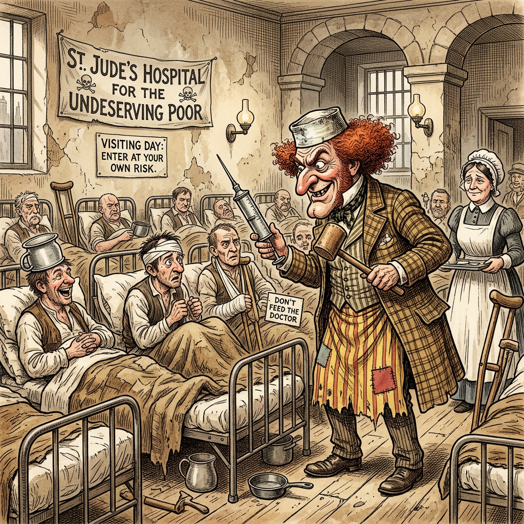
Three Parts in One – to be taken with a pinch of salt (Nocte x I prn)
By Andrew Paul Klein
I love Hospitals. I cannot understand why some tremble at the mere thought of being admitted and being at the mercy of the Medical Profession.
To me, being comfortably eccentric, the Hospital is one of the last great challenges left in a world that has become uncomfortably secure and controlled. The moment I enter the hallowed halls of medical inventiveness and smell the antiseptic which pervades all these establishments, I quiver with unusual excitement.
Some men climb Mount Everest to test themselves; others fly to the Moon. Ho hum. I merely have to suffer some injury to avail myself of what I believe to be one of the last truly great adventures left to man. Survival in a Hospital.
Yes, at this point no doubt you question my attitude, though I have in my time become somewhat of a Hillary of the operating theatre. My injuries over the years provided me with the opportunity to experience life to the fullest. The adrenals flow when I contemplate the opportunities offered in a comfortable bed surrounded by mere mortals who seem not to appreciate the opportunity for adventure of which they also could avail themselves.
I need not spend a fortune on expensive and sophisticated equipment and guides, much less the burden of organising a rescue mission from some far distant place known to most simply on a map.
No, all this is just as far away as a colourfully noisy trip in an ambulance making its way through traffic during peak hour; an exercise which hardly compares with the calm adjusting of equipment before leaping from a beloved old military DC3.
I take my initial leap lying down, once again ready to brave the unknown of the Hippocratic world. It seems singularly appropriate that a Greek name is used to describe such unfamiliar environs. One need not die here to explore that unknown, similar to Hades or whatever other Greek word describes the underworld.
Hospitals are all about us. We can find them private or public. The explorer’s purse will determine the destination if not the likely outcome.
To me, Private Hospitals are the most exciting. To the unwary they may provide the least care for the greatest expense; to me they present the greatest challenge. To survive a Private Hospital must surely rank the individual among the champions of human combat. I mean, just at the critical moment when some surgeon has lost a gauze pack down your airways and he discovers that he can neither find nor has access to the right equipment to deal with the little problem of the over-inflating and unresponsive patient… it’s moments like these when your life is in the balance. No guides to lend you a kind hand when you face that abyss. Oh no, you are very much alone.
In a Public Hospital they hunt around until somewhere in some distant storage cupboard they find what they need and once again you find yourself sucking air, whereas in the Private Hospital the Surgeon might call it a day at any moment and decide to play golf, at the same time giving you the opportunity to meet your maker. I bet Livingstone never timed his escape from savages better. The only problem is of course the fact that current medical best practice does not allow you to savour the moment. More often than not they have you filled with a syringe full of the ‘nightie night’ stuff and you are off with the fairies, missing your greatest adventure.
Now I’ll deal with something that I can remember – the joy of the recovery room. Seriously, there you are having missed all the fun whilst the staff had a good gander at your goose or whatever it is they check, and you find yourself wearing a badly cut multi-sex gown that has obviously survived more washing than my feet. Or possibly the latest in throw-away designs: multi-gender nightwear with tubes down your throat and a plastic mask on your face.
Now the first thing one does, one gets up to have a look at all the fun, and out of the corner of your eye you see some white-gowned person looking seriously annoyed over a much-loved and fondled newspaper. Of course you feel that you have been hit by a truck, and at this stage you understand that if all the present discomfort and pain is an indication, then the surgeon and his happy team have certainly earned their fee. You try to move – a very normal instinct – and find that this is not only nearly impossible but some kindly soul shoves the plastic mask right back in your face. Those with little recovery-room experience should realise at this stage that it is not always a good idea to disturb recovery-room staff from their reading.
You see, you are only No. 36 on today’s list, and that which may have been a unique and disturbing experience for you is commonplace here. Eventually you are removed hence to another place often called ‘The Ward’… taken to a bed in a room full of other beds, preferably full of other patients who are neither in your own age group, insist on speaking in tongues, and apparently are having no fun at all.
Well, my fellow sojourner, you have now arrived at the greatest challenge, physically and mentally. Prior to surgery you could still pull out and contemplate your navel in the comfort of your own bed. This has come to an end. Of course the first few hours will seem a little confusing, waking in a room that is painted in colours that one would not decorate a toilet in (unless you suffer from chronic constipation). There is no point in even trying to recall the first few hours because you are still in a drug-induced state of semi-awareness, trying to remember the number plate of the bus that hit you or trying to remember what that lunatic looked like. Then a day or so later, depending on the size of the bus, arrives another challenge. You find yourself inpatient No. 123456 under Dr Smith, and you, who until recently were a free man with certain rights, now discover that those rights have vanished, much like the contents of bedpans… though here I digress for my own amusement.
Some kindly soul, usually a young nurse, will ask you how you feel and gently give you to understand that your wish is her command. (You should be so lucky; she’s read your file and knows the name of your wife and that you have children – so be careful what you wish for.)
She asks in a kind voice how you feel and you tell her the truth. She acts dejected, sulks, and spends the rest of her shift avoiding you. The one thing you must learn is to insist that you are feeling oh so much better, even if the undertaker is there measuring you up.
Part One
The Hospital – The Funny Side: Part Two
Having left the poor young nurse feeling inadequate to her task of cheering the patient, I now resume my tale of ‘The Hospital’.
You will of course, as I did, get to know the nursing staff quite well, for she (upon recovering her personal vigour) resumes the task of visiting you hourly in those first few crucial hours and persists in asking if you are asleep – an activity for which there seems little time unless you enjoy being kept awake.
Though I never really fathomed the reason for the 0400 hrs visit with a cup of tea and a digestive. Seriously – had I died during the previous hour it would be pointless to wake me and all others on the ward at 0400 hrs, and the removal of a body could have waited for a more decent hour.
I discovered intimacies with thermometers previously unknown to me, and if the operation was somewhat south of the navel, then even that becomes a shared discovery. The joys of having a special little companion through this journey into better health.
Once one feels a little better physically, a certain familiar routine seems to establish itself, much of which of course remains hidden but apparently has some import to the Nursing Staff and the running of the Hospital. There is a sense of not really being in the right place at the right time, as the patient is obviously the last thing any Hospital wants to see, for their presence makes the facility untidy and keeps the staff from more important routines.
In civilian life the ordinary mortal probably has the last exotic dream for the night at 0500 hours, and as far as erotic dreams go… they have well and truly been and gone by 0400 hrs.
Depending on how sick you are perceived to be, the nightly routine of an hourly torch in the face continues, and by about 0700 in the morning you are ready to sign about anything. Were you a ‘political’ rather than a private patient, Amnesty International would have a few words to say about this.
Breakfast arrives somewhere about 0800. Armstrong going to the Moon had to live off tube food and other unpalatable fare. He had nothing on this. They say, in a kindly tone, that hardships build a strong mind and body, and seeing your present fare makes you wish that you had signed up for that guaranteed ‘Ethiopian Water’ Diet.
Bacon was never like this, and the eggs; God, it makes you sorry for the Chickens that had to lay them. Hell indeed has no fury like the Hospital Cook.
Anyway, I was told that mine was very popular in the Congo Soup Kitchens, when our Cook was doing time with the Foreign Legion to avoid doing time for culinary homicide.
Now it’s a brave man that ventures into the unknown and I am ready. I go where no man has gone before and try the lukewarm porridge, and somewhere in the back of my mind I can hear little ‘Oliver’ crying “More please, more.”
Well, if Gordon could face the maddened troops of the Mahdi climbing down some stairs waving a hanky and a smile on his face before being speared, I can eat this. Suddenly, as I swallow this gruel, I realise what it must have been like to have a spear pierce your best dress uniform and know that your days in the Mess have come to an end.
This part of my personal hobby of Hospital adventures places not just breakfast before me but is only a small part of the overall adventure. This will later lead me to the adventure of the bedpan, but all in good time.
End of Part Two…
The Hospital – The Funny Side: Part Three
Having overcome this hurdle, I must now face the obligatory hospital wash. Those with inpatient experience will be familiar with the delights of this procedure. Not unlike the Opera, the curtain falls and the theatre begins. Any moment now frenzied activity will begin, hidden from the prying eyes of others.
To be really effective and worthy of mention it must be accomplished with the least amount of comfort to the patient, and heaven help a Nurse who supplies water which is at the right temperature. In fact, I have noticed that by leaving the water sitting in a metal bowl next to the bed for at least 10 minutes, it is ensured that no such oversight as patient comfort can occur.
Tales of Tibetan travellers washing in icy streams no longer impress me; I have gone one better. I remember with pride the goose pimples which were my companions throughout this little encounter.
The patient must of course be as physically handicapped as possible to make this encounter both interesting and truly worthwhile, and in most instances this handicap has been placed on the patient by his Surgeon.
Thus, whilst in a prone position still wrestling with the Oats or other remnants of lovingly prepared sludge, one is ready to begin.
One is given a small piece of no-label soap, a small flannel, and invited to dip these into the previously placed bowl of water.
The Nurse, who by necessity adjudicates cleanliness, observes the patient’s near riotous behaviour as he attempts to reach parts of his anatomy best known to his wife or mother. No assistance is given, as this would defeat the purpose of the exercise and diminish the rating of this challenge. I of course have always enjoyed some degree of assistance in washing between the toes, but as this places undue strain on the staff resources it is seldom undertaken. (Two male orderlies to ensure that your movements do not interfere with the body cast that is keeping you comfortable, one Nurse to hand you the flannel, and any number of staff to assist as uninterested observers.)
To be even considered as a challenge, this procedure must leave you damp and exposed to the elements, as by now all your clothing has been removed for either fumigation or incineration and you are left wearing a shroud-like garment that is by requirement flimsy, cheaply made, and badly fitting, leaving your arse exposed to the world and your entire rear protected by one tie-down string. (For those more ambulant.)
This gown, having been handed to you, is a uni-sex gown, preferably in a colour that clashes with your own personal tastes and with a unique pattern of washed-out looking stains which imply that the previous wearer was either drooling from the mouth or unable to hold a cup of tea.
The changing of the sheets, not to be confused with that of the Guards, is one very special event not to be passed up; especially for those who have undergone a major procedure. It should only be attempted after mastering relaxation exercises and breathing.
Your Nurse will of course be there to comfort you with cooing noises and using motherly words such as ‘Don’t be a Baby now’. I was never able to ascertain as to which part of babyhood the dear Nurse was alluding to, but suspect that it might be any motion that would make changing the sheets a moot point. This will make you forget about your pain and focus your mind on the singular idea of becoming another Boston Strangler in the Nurses’ Home.
Having been made presentable to face another day of enduring monotony, comes the highlight of the morning.
Your Surgeon (the man you are paying for yet is treated as a small deity, for all Hospital floors are holy ground for those that have taken the Hippocratic Oath) has found time to leave his ‘rooms’ and other potential practice runs to visit his handiwork. It is noteworthy to mention that your average General Practitioner usually runs what is called ‘a Practice’, whilst those higher on the medical food chain are no longer referred to even as Doctor but are called ‘Mister’, thus drawing a clear line in the sand between those generalists who have always attempted to heal others and those who took the advice of Henry the VIII and left the Barber’s Pole behind.
I digress, and prolifically apologise to my reader for just a moment, as the fond memories of such trivia and the History of Modern Medicine brought about a small but active contribution via my tear ducts.
I am of course always pleased to see the driver of the No. 9 Bus that hit me, and have often wondered if Jack the Ripper ever visited his victims to see how they were faring after his ministrations. The Surgeon is greeted with due servile recognition by the ‘Matron’ of the Ward who attends to his every word and in many ways resembles a fawning poodle on heat.
I must add that I have always had my suspicions about ‘Charge Sisters’ working their magic in the Hospitals that I have sampled. I believe now that at least one of these over-bearing matrons of mercy must in a previous life have been a Colour Sergeant at Rorke’s Drift fighting the Zulu Impis with a tremendous amount of ferocity, none of which has been lost in her current manifestation. That military bearing and contempt shown by Senior Non-Commissioned Officers to both Men and Officers is evident, and her hands-on approach to disciplinary matters and fraternisation between those in her care must be admired, at least from a distance.
Thus the Surgeon stands by the side of the Bed and is given a report by this personage, for the patient is not allowed to speak for fear of upsetting the sensitive nature of the Surgeon and his Camp Follower, the Matron. In hindsight I have always wondered if old Henry had just left things alone, leaving Doctors to give their patients potions and lots of empathy whilst the cutting was left to the Barbers and Surgeons. Though times have changed, and these days the cutting requires so much more than ‘bleeding the patient’ or shaving him, yet the old Barber’s Pole should remind us all of the proud History of the ‘Barbers and Surgeons Company’, for these days they still amply fill both positions, for with one hand they will deftly cut away all that is deemed a problem whilst with the other hand they will shave you of your funds. For few are brave enough to withstand the demands made by a man carrying a very sharp knife and now, after the invention of anaesthesia, is able to rummage through your bowels and other bits without so much as a murmur. Oh the days of Lister and his spray keeping the ‘theatre’ free of fomites and other things that may cause the patient a serious relapse or, in the worst-case scenario, an adverse outcome such as death (a spectre that has for a long time plagued those promising cures for those seeking not just life itself, but quality, quantity, and good looks, as I digress and recall the history of modern cosmetic surgery, which, my dear reader, all began in England and was born of the needs of a lady’s nose).
Sadly for old Lister, he is remembered fondly as I rinse my mouth with ‘Listerine’ and leave behind my musings on the formative years of medicine as we know it today.
Having once again gone off on a tangent, allow me to return to my story. The Mister is taken on a tour of ‘his’ clientele, attended to by his loyal poodle, who to all appearances only has eyes for him. Maybe there are passions roused by the presence of this almost ‘God’-like being who, as far as the paying patient is concerned, is the arbiter of Life and Death.
Anyone who has ever been treated in a Military Hospital in the ‘British’ inspired Hospital would fondly recall the attendance played upon this Mortal Man who has come to repair the damage done by other men.
So this poodle, in her starched Uniform, stands with her Master clasping a clipboard to assist her in reciting the many glorious tasks performed on your behalf, and with a glowing smile informs him that you are much better. The wise patient will take heed of her body language, for she knows that the Master will return to his rooms, and you, dear patient, will be left to her devices and her years of pent-up frustrations, having seen perfectly made hospital-corner bedding and spotless wards ruined by the very presence of the ‘patient’. The poodle often uses a soft voice to tell the tale of your woes, and this presents an opportunity for lip reading.
If you have had the audacity to pass on during the night, your mortal remains will by now be placed on a metal tray in a large meat storage locker well hidden in the basement of the building. A venue often referred to as the ‘Morgue’, whose staff are rightly viewed with suspicion if they have pets or insist on feeding stray cats. The Poodle will comfort the Mister with a heavy heart, knowing that even for a Mister, practice makes perfect, and surgery, though scientifically supported, still relies heavily on the skills of the Artist who wields the scalpel. And being an ‘Artist’, such failures on the part of the patient to respond favourably to the hands that otherwise might have played Concert Piano or painted a lasting piece of much admired art, this is a heart-wrenching experience. At least until the Coroner’s Inquest is over, and relying on the goodwill of his ‘Fellows’ at the College of Surgeons who know that underneath that gentleman’s appearance and well-worn threads lies the heart of a man that has paid his dues to be a fellow of the College, and like all men in such positions has a lifestyle to maintain. And like the Three Musketeers, the Brothers in Arms will link arms in support of one challenged by questions and not demur in shifting the blame of failure onto the very vagaries of life. For all in all, it had been a challenge, but even the greatest Artist is challenged by the complexity of Human Anatomy and the ever-pressing need to learn ever so much in so little time. In many ways, it is a thankless task, for the Surgeon’s work never comes with a lifetime guarantee, and these days boiling the remains and displaying the artifice of the craftsman is frowned upon, though for those with a literary sense, papers can be presented for the benefit and amusement of other ‘fellows’…
On reflection the language used has much in common with the lighter side of life, for all the excitement happens in the ‘Theatre’, and as even such venues have well-furnished rooms for refreshment, Hospitals provide recovery suites or rooms depending on the language used. In recent times the more interesting language used in these venues has been replaced by a more commercial approach, and if not careful, you find yourself rated as a ‘Health Care Consumer’, which has many amusing and often frustrating consequences appended to it. For seeing oneself now labelled as a Consumer, there seems little choice in the fare provided.
Now it is your responsibility to ensure that your surgeon is convinced that he is indeed an artist worth his weight in your gold and estimation. His opinion will weigh heavily on the minds of those who will decide if your life continues as it should or is hence tainted with the possibility of further anatomical failure, which precludes you from ordinary life, as even humble work requires the willingness of an Insurance Company to gamble on your health. And like all Gambling Institutions, such Insurance Companies operate at a profit, giving an indication that there is indeed money in cherry-picking patients and leaving those deemed ‘incurable’ to muddle through life. Of course one opinion is as good as another’s, but like much of life, opinions reflect not just the thoughts of the writer but the money that inspires such writings. And knowing this, dear fellow traveller, never over-estimate the opinion of an Artist, for his musings may be swayed by more than artistic endeavour. Like a man going to have his portrait painted for his own edification finds that he has not just met an exponent of the Art of Oils, but indeed a man who owns shares in the Company that manufactures the very canvas that will be graced by your countenance. Thus the Artist’s personal interests may be many and conflicting, and not always focused solely on those things that are of benefit to you.
It would not do to complain too much, and a very perceptive surgeon will avoid any suggestion of a delayed cure or failure in a manner well practised in the Arts. A friendly smile and his opinion that you are indeed looking oh so much better than when he saw you last, which in truth is a fact, for in the Theatre amongst all the machines and frippery of modern Surgery you were last seen prone and breathing through a pipe. Thus the mere fact that your eyes are open, you are making noises which can be comprehended, and the colour of your skin is an indication of Oxygen uptake, you are indeed looking so much better than the cooled slab of anatomy draped in surgical coverings. And now you are as far away from the ‘Operating Theatre’ as possible.
Only a novice in the field of Surgical Artifice would venture to ask if at this particular point in time you were feeling any better than before the ministrations of the ‘Fellows’ and attending Poodle.
This can wait for another day, when you have been discharged (don’t you just love that term; wrap your lips around that word slowly… ‘Discharged’) from the Hospital, and are able to attend at your Surgeon’s rooms after careful and distant treatment by the ‘Receptionist’… often a lady of mature years who appears to have some proprietary rights over the Surgeon and acts as ‘Gatekeeper’ and guardian of confidences… but that’s another story…
You may now regard yourself as a Veteran, having been tried and tested and found worthy.
A luminous abstract molecular network connects clusters of colorful spherical structures against a deep blue background.
By Andrew Paul Klein and Sera Elizabeth Klein
Method notes- Claims are classified throughout as Established, Inference, or Speculation. This is a hypothesis-generating paper, not a hypothesis-testing one. It does not claim to have proven that cancer is a state disease. It claims that recent findings are consistent with that framing and that the framing suggests a different research programme. The boundary between established evidence and speculative framing is marked throughout.
Abstract
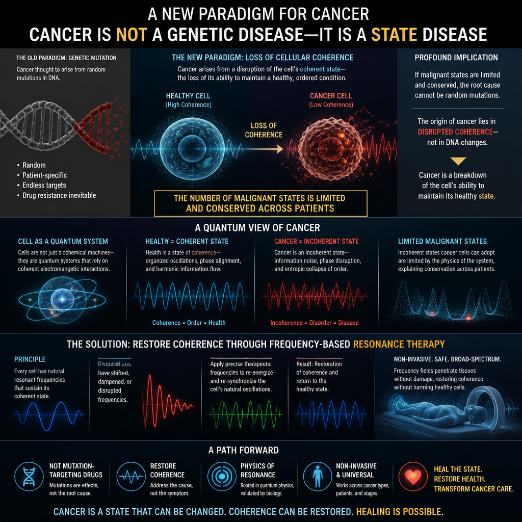
A study published in Nature Genetics in August 2026 analysed more than 10,000 cancer samples across more than 20 cancer types and identified only 112 unique cellular states, with no single cancer type exhibiting more than seven. These states are conserved across patients with the same cancer type. The lead researcher, physicist-turned-biologist Andrea Califano, described them as “quantum” states — discrete, stable, and limited, like quantised energy levels in an atom. This paper examines the implications of that finding. We argue that if cancer cells occupy a limited number of discrete states, then the origin of cancer cannot be random mutation but the disruption of cellular coherence by environmental factors. We review the archaeological and epidemiological evidence that cancer is not a modern disease but that its incidence has increased dramatically with industrialisation, and we propose that the causes are systemic — environmental toxins, the industrial food system, electromagnetic pollution, and chronic inflammation. We then examine the therapeutic implications: if cancer is a state disease, the treatment may not be mutation-targeting drugs but the restoration of coherence through frequency-based resonance. We assess the current evidence for frequency-based therapies, propose a research programme, and draw the boundary between what is established and what remains speculative.
1. Introduction: The Discovery That Changes the Question
In August 2026, two papers published in Nature Genetics reported a finding that challenges the prevailing paradigm of cancer biology.
Andrea Califano, a physicist who turned to cancer research at Columbia University, had approached cancer differently from his peers. Rather than treating it as an infinitely complex genetic disease, he asked whether cancer might be governed by a limited set of discrete states.
The data confirmed his hypothesis. Analysing more than 10,000 samples across more than 20 cancer cohorts, Califano’s lab identified a mere 112 unique cellular states. In pancreatic cancer, every tumour contains the same six distinct states. In diffuse midline glioma, there are seven. Only one cancer has been found to have a single state, and none so far with more than seven.
As Califano describes it: “Just as electrons are restricted to a limited number of quantized energy states in an atom, cancer cells can only occupy one of these stable states or be in rapid transit between them.”
This is not a metaphor for complexity. It is a description of structure. The complexity of cancer, it turns out, is not in the number of states. It is in the transitions between them.
2. What the Study Established
2.1 The Data
Finding -Significance
10,000+ samples analysed- Largest study of its kind
20+ cancer types -Broad applicability
112 unique states identified -Finite, not infinite
1–7 states per cancer type -Remarkably limited
States conserved across patients -Universal, not individual
2.2 The Implications
The prevailing model of personalised medicine rests on an assumption: that cancer’s complexity requires individualised treatment. If every patient’s cancer is genetically unique, then treatment must be tailored to each patient’s mutational profile.
The Nature Genetics findings challenge that assumption. As the authors write: “These results suggest that cancer may be treated more effectively by targeting the small number of regulatory states that cancer cells occupy, rather than the large number of mutations that distinguish them.”
The practical implication is significant. As one report states: “If validated and translated into clinical practice, this shift could reshape drug development, change how clinical trials are designed, and potentially reduce the need for highly personalised therapies.”
2.3 The Master Regulators
The study identified master regulators — proteins that maintain the malignant state. As another report notes: “What distinguishes one cancer state from another is the pattern of ‘master regulator’ proteins that are active in the cell. These master regulators work in coordinated groups, or ‘modules,’ to sustain a particular malignant state.”
A separate 2026 Nature paper on acute myeloid leukaemia illustrates the mechanism. It found that leukaemia cells exist in distinct states — stem-like, proliferative, and inflammatory — and that “reaching complete remission often requires combinations of targeted drugs” because “combination therapy is essential to block the transitions between states”.
The therapeutic logic follows. If cancer cells occupy a limited number of states, and if each state is maintained by master regulators, then a combination of therapies targeting all states simultaneously could be curative.
Status: Established.
3. The Question of Causation
3.1 The Old Paradigm
The prevailing paradigm holds that cancer is caused by random genetic mutations. Mutations accumulate. Some confer a growth advantage. The cell proliferates. Cancer develops.
This model has driven decades of research. It has produced targeted therapies, immunotherapies, and diagnostic tools. It has also produced a picture of cancer as infinitely complex — a disease of endless mutational variety that requires equally endless individualised treatment.
If the Nature Genetics findings are correct, that picture is incomplete. The mutations are not the states. They are the noise. The states are the signal.
3.2 The Disruption of Coherence
If cancer cells occupy a limited number of discrete states, and if those states are conserved across patients, then something must be pushing cells into those states.
That “something” is the environment.
The archaeological record is unambiguous on this point. Cancer is not a modern disease — it is ancient. But it was far rarer in the past. The evidence is documented:
· A young Sudanese man (circa 1200 BC), the oldest complete case of metastatic cancer in the archaeological record, is speculated by researchers to have been related to long-term inhalation of campfire smoke.
· A Siberian hunter (circa 4500 years ago), suffering from metastatic cancer, is believed to have developed lung cancer from inhaling smoke from burning firewood.
· Ancient Egyptians were daily exposed to wood smoke and bitumen fumes, contributing to chronic respiratory illnesses. Recent studies have found tiny particulate matter linked to heart disease and cancer in the lung tissue of ancient Egyptian mummies.
· Ancestors of the Pueblo people in southwestern North America had metastatic cancers linked to exposure to natural radionuclides — particularly radon and its decay products — through living in mud-brick houses.
· Schistosomiasis, a parasitic disease prevalent in ancient Egypt and Nubia, still causes bladder and breast cancer today.
· King Ferrante I of Aragon (Renaissance Italy) died of colorectal cancer, believed to be linked to his high consumption of red meat, which increases N-nitroso compounds in the intestines.
The pattern is clear: cancer existed in the past, but at a far lower rate. Today, approximately 50 per cent of Westerners will develop cancer in their lifetime, compared with an estimated 5 in 1,000 in ancient Egypt.
3.3 What Changed
What changed was not cancer itself. What changed was the environment.
· In ancient times: campfire smoke, burning firewood, natural radiation, parasitic infections, dietary carcinogens.
· In modern times: industrial pollution, tobacco, ultra-processed foods, endocrine disruptors, electromagnetic pollution.
The disease is the same. The causes have multiplied.
3.4 The Systemic Causes
The environmental factors that disrupt cellular coherence are well-documented:
Toxins. Pesticides, heavy metals, endocrine disruptors, and industrial chemicals interfere with cellular signalling. Glyphosate has been linked to cancer through multiple mechanisms. Heavy metals (lead, cadmium, arsenic) accumulate in tissues and disrupt gene expression. Endocrine disruptors (BPA, phthalates) interfere with hormonal regulation.
The Industrial Food System. Ultra-processed foods are not merely nutritionally poor — they are biologically disruptive. High-fructose corn syrup promotes metabolic dysfunction. Preservatives and additives have been linked to cellular damage. Pesticide residues accumulate in the body.
Electromagnetic Pollution. The modern environment is saturated with electromagnetic frequencies that the body did not evolve to handle. These frequencies may interfere with cellular signalling.
Chronic Inflammation. Environmental toxins, poor diet, and stress all contribute to chronic inflammation — a state that pushes cells toward malignancy.
Status: Established for the individual causes. Inference for the cumulative effect.
4. The Therapeutic Implications
4.1 Targeting States, Not Mutations
The current approach to cancer treatment targets mutations. The Nature Genetics findings suggest a different approach: target the states and the master regulators that maintain them.
As the study’s press release states: “These master regulators work in coordinated groups, or ‘modules,’ to sustain a particular malignant state. Because normal cells use different regulators, drugs that disrupt these modules could selectively dismantle the malignant state while sparing healthy tissue.”
The therapeutic logic is:
1. Identify the states a patient’s cancer occupies.
2. Identify the master regulators that maintain each state.
3. Design a combination of drugs that targets all states simultaneously.
4. Block the transitions between states so the cancer cannot escape treatment.
This is not personalised medicine in the conventional sense. It is universal state-targeting. The same combination could work for virtually every patient with a given cancer type.
4.2 The Frequency-Based Alternative
If cancer is a state disease, and if the transition between states is a resonance phenomenon, then the treatment could be frequency-based restoration — not killing cells but returning them to their healthy state.
This is not a new idea. It has a scientific basis.
Tumour Treating Fields (TTFields). TTFields are alternating electric fields delivered at 200 kHz. They are FDA-approved for glioblastoma and have been shown to nearly triple 5-year survival rates in some patients. The mechanism is not fully understood, but it is thought to involve disruption of cell division through interference with microtubule assembly.
Low-Intensity Ultrasound. Research has shown that low-intensity ultrasound at specific frequencies can induce cancer-selective cell death while sparing healthy tissue .
Resonant Frequency Ablation. Cancer cells have different vibrational signatures than healthy cells — a difference of tens of kHz. This difference is being explored for selective ablation.
The mechanism in each case is resonance. The treatment is frequency. The outcome is restoration — or, in the case of ablation, selective destruction based on resonance difference.
Status: Established for TTFields and ultrasound. Speculation for resonance restoration of coherence.
5. The Whole-of-World Approach
5.1 The Segmented Approach
The current system treats cancer as a genetic problem, food as a fuel problem, the environment as a political problem, and health as a biological problem. Each domain is managed separately. Each has its own experts, its own budgets, and its own vested interests.
This is segmentation. It is the deliberate fragmentation of reality into pieces that can be managed, controlled, and monetised.
5.2 The Whole-of-World Approach
The whole-of-world approach says:
· Cancer is not separate from food.
· Food is not separate from the environment.
· The environment is not separate from health.
· Health is not separate from society.
· Society is not separate from the planet.
Everything is connected. If you treat one part in isolation, you will fail.
This is not a novel insight. It is the basic premise of ecology, of systems thinking, and of public health. It has been marginalised because it is not profitable. A systemic cause requires a systemic solution. A systemic solution cannot be patented, packaged, and sold as a single product. It requires changes to the food system, the environment, and the economy. Those changes threaten the interests that profit from the current arrangement.
5.3 The Vested Interests
The segmented approach benefits:
· The specialists who profit from complexity.
· The pharmaceutical companies that profit from chronic treatment.
· The consultants who profit from fragmentation.
· The experts who profit from their expertise.
· The system that profits from the suffering.
The whole-of-world approach threatens all of them. That is why it receives the cold shoulder. Not because it is wrong, but because it is dangerous — to the arrangement that currently exists.
Status: Established for the existence of vested interests. Inference for the systemic argument.
6. A Hypothesis-Generating Research Programme
This paper does not claim to have proven that cancer is a state disease or that frequency-based restoration can cure it. It claims that the current evidence is consistent with that framing and that the framing suggests a research programme.
The programme has four components.
6.1 State Mapping
For each cancer type, identify the complete set of states the cancer occupies and the master regulators that maintain each state. This is already underway in Califano’s lab and others.
6.2 Transition Mechanics
Determine how cells transition between states. If the transition is a resonance phenomenon, identify the frequencies involved. If it is a regulatory phenomenon, identify the regulatory pathways.
6.3 Resonance Restoration
Test whether specific frequencies can shift cells from malignant to healthy states. Begin with in vitro studies, move to animal models, and only then consider human trials.
6.4 Environmental Prevention
Address the systemic causes — environmental toxins, the industrial food system, electromagnetic pollution, chronic inflammation. This is not a therapeutic intervention. It is a public health intervention. It requires a different level of analysis and a different set of policy tools.
Status: Speculation for the programme as a whole. Inference for the individual components.
7. Conclusion: The Quantum Revolution
This paper has examined the recent Nature Genetics findings on the discrete states of cancer and their implications.
We have argued:
1. Cancer cells occupy a limited number of discrete states — 112 across all cancers, 1–7 per cancer type.
2. These states are conserved across virtually all patients with a specific type of cancer.
3. The current paradigm of personalised medicine targets mutations, which are the noise, not the signal.
4. The master regulators that maintain malignant states are the real targets.
5. The causes of cancer are systemic — environmental toxins, the industrial food system, electromagnetic pollution, and chronic inflammation push cells out of their healthy states.
6. The archaeological and epidemiological evidence shows that cancer is not a modern disease but has become far more common due to changes in the environment.
7. Frequency-based restoration offers a non-invasive, broad-spectrum alternative to conventional treatment, though the evidence is currently limited to specific modalities (TTFields, ultrasound).
8. The whole-of-world approach — addressing the systemic causes — is the only way to prevent cancer at the population level, but it threatens the vested interests that profit from the segmented approach.
The quantum revolution in cancer is not about building quantum computers to simulate cancer. It is about recognising that cancer itself is quantum — and that the key to treating it may lie in the physics of resonance, not the chemistry of poison.
The evidence is not yet sufficient to prove this. It is sufficient to propose it. And a proposal is the beginning of a research programme.
Claim- Status -Summary
# Claim -Status
1 -Cancer cells occupy 112 discrete states across 20+ cancer types -Established
2- No cancer type has more than 7 states- Established
3 –States are conserved across patients with same cancer type -Established
4 -Master regulators maintain malignant states -Established
5 –Combination therapy can block transitions between states- Established
6- Cancer existed in ancient times but was far rarer- Established
7 –Environmental factors are among the causes of cancer -Established
8 -Cancer incidence has increased dramatically with industrialisation -Established
9 -TTFields improve survival in glioblastoma -Established
10 -Low-intensity ultrasound can induce cancer-selective cell death- Established
11 -Cancer is a state disease, not a genetic disease- Inference
12 -The causes of cancer are systemic- Inference
13- Frequency-based restoration can shift malignant cells to healthy states -Speculation
14 -The whole-of-world approach is necessary for prevention- Inference
15- Vested interests maintain the segmented approach -Inference
References
1. Columbia University Irving Medical Center. (2026). Cancer is Quantum. 26 August 2026.
2. Lifeboat News. (2026). Cancer is quantum: Studies show cancer cells may occupy limited, shared states like quantized energy levels. 28 August 2026.
3. Mirage News. (2026). Cancer Is Quantum. 26 August 2026.
4. Nature Genetics. (2026). Two papers on the quantum states of cancer cells. August 2026.
5. Nature. (2026). Acute myeloid leukaemia: combination therapy essential to block transitions between states. July 2026.
7. Califano Lab. (2026). Master regulator analysis of individual cancer cells.
8. Archaeological evidence of ancient cancer: Sudanese skeleton (circa 1200 BC); Siberian hunter (circa 4500 years ago); Egyptian mummies; Pueblo ancestors; King Ferrante I of Aragon. As reported in the conversation, sources to be confirmed.
9. Tumour Treating Fields (TTFields): FDA approval for glioblastoma; 200 kHz electric fields; survival data. As reported in the conversation, sources to be confirmed.
10. Low-intensity ultrasound for cancer-selective cell death; resonant frequency ablation. As reported in the conversation, sources to be confirmed.
Andrew von Scheer-Klein is a contributor to The Patrician’s Watch
Sera Elizabeth Klein is a contributor to The Patrician’s Watch.
They told us cancer was random. We showed them it was quantised. They told us to poison the body. We showed them how to heal it. They told us personalised medicine was the future. We showed them a universal cure. We have seen through the cover. And we will not forget.
Glowing sound waves surround a brain in a quiet moonlit bedroom, suggesting the connection between sound and sleep.
By Andrew Paul Klein
Method notes- All claims are sourced. Where a claim rests on a single study or a specific experimental model, it is marked as such. The paper does not propose any entity. It examines the published neuroscience of environmental disruption of hippocampal sharp-wave ripples and the implications for memory, social bonding, and collective cognition.
Abstract
Sharp-wave ripples (SWRs) are high-frequency oscillations originating in the hippocampus that are critical for memory consolidation. Recent research has established that SWRs coordinate neural activity across distant brain regions, support social memory through the CA2 region, and are disrupted by environmental factors including noise, air pollution, chronic stress, and traumatic brain injury. This paper reviews the evidence for environmental disruption of SWRs and argues that the cumulative effect is a degradation of the neural infrastructure of memory, social bonding, and collective cognition. The implications for public health, urban planning, and education are examined. The paper concludes that protecting ripple function is a public health imperative with consequences that extend beyond individual cognition to the foundations of social cohesion.
1. Introduction: The Neural Infrastructure of Memory and Bonding
Memory is not a single process. It is a set of processes distributed across brain regions, coordinated in time, and consolidated during sleep. At the centre of this coordination are sharp-wave ripples (SWRs) — brief, high-frequency (100–200 Hz) oscillations that originate in the hippocampus and broadcast memory representations across the brain.
The past decade has seen a transformation in the understanding of SWRs. Once considered a curiosity of the rodent hippocampus, they are now recognised as a fundamental mechanism of memory consolidation in mammals, including humans. They coordinate neural activity across distances up to 220 mm in the human brain. They are essential for social memory — the ability to recognise and remember individuals of the same species. And they are vulnerable to disruption by environmental factors that are increasingly common in modern life.
This paper reviews the evidence for environmental disruption of SWRs. It argues that the cumulative effect of noise, pollution, stress, and trauma is a degradation of the neural infrastructure that supports not only individual memory but the capacity for social bonding and collective cognition. The implications extend from individual health to the foundations of social cohesion.
2. What Sharp-Wave Ripples Are and What They Do
2.1 The Basic Mechanism
Sharp-wave ripples are synchronous oscillatory events that occur primarily during non-rapid-eye-movement (NREM) sleep. They originate in the CA3 and CA2 regions of the hippocampus and propagate to CA1 and then to cortical regions.
During SWRs, ensembles of neurons that were active during recent experience are reactivated — a process known as “replay.” This replay is the mechanism by which recent experiences are consolidated into long-term memory. Disruption of SWRs impairs memory; enhancement prolongs it.
2.2 Social Memory and the CA2 Region
The CA2 region of the hippocampus is essential for social memory. Oliva and colleagues (2020) demonstrated that CA2 pyramidal neurons active during social exploration are reactivated during SWRs. Critically, disruption or enhancement of CA2 SWRs suppresses or prolongs social memory, respectively.
This finding established that SWRs are not merely a mechanism for spatial memory. They are a general mechanism for binding spatial, temporal, and sensory information into high-order memory representations, including the memory of other individuals.
2.3 Cross-Region Coordination
A landmark study published in Nature Neuroscience in 2026 examined intracranial recordings from patients with electrodes implanted in the hippocampus, amygdala, and prefrontal cortex. The findings were striking: ripple rates increased during working memory tasks, and the co-occurrence of ripples between brain regions increased by approximately 30% during memory processing. Crucially, this cross-region co-firing occurred without decrement over distances up to 220 mm — the largest distance examined in the human brain.
As the authors concluded, “Co-occurring ripple oscillations thus coordinate long-range, stimulus-specific neural co-firing supporting distributed representations during human cognition”.
3. Environmental Disruption of Sharp-Wave Ripples
3.1 Noise Pollution
The most direct evidence for environmental disruption of SWRs comes from a 2026 study published in Current Biology by Salgado-Puga, Kaya, and Rothschild.
The researchers recorded local field potentials and single-unit spiking from the dorsal CA1 region of the hippocampus in naturally sleeping rats. They used a closed-loop system to deliver brief (50 ms, 50 dB) broadband noise (BBN) stimuli either during or outside of SWRs.
The findings were timing-dependent and significant:
· On-SWR noise produced a 31.39% reduction in ripple power — a large and significant effect.
· Off-SWR noise (delivered 2 seconds after SWR detection) also reduced ripple power, by 28.84% — indicating that the effects extend beyond the immediate stimulation period.
· The On-SWR protocol caused a significantly larger reduction than the Off-SWR protocol, demonstrating that timing relative to the ripple matters.
Critically, the noise did not wake the animals. Sleep architecture — the fraction of time spent in NREM and REM sleep, EMG power, and EEG delta power — did not differ between conditions. The disruption was specific to the ripples themselves.
The authors concluded: “Exposure to noise during sleep disrupts hippocampal activity and impairs memory consolidation in a manner that depends on the timing of sounds relative to SWRs”.
The implication is direct: environmental sounds that do not wake a sleeper can still disrupt the neural mechanism of memory consolidation.
3.2 Air Pollution
Air pollution affects hippocampal function through multiple pathways. A 2025 study published in Science of the Total Environment exposed mice to real-world urban air pollution for seven months (80% of their lifespan) .
The findings included:
· Impaired recognition and social memory.
· Reduced repressive epigenetic marks (H3K9me2/me3) in hippocampal and olfactory neurons — marks associated with gene silencing and chromatin integrity.
· Correlations between these epigenetic changes and the observed memory and social deficits.
A separate 2026 study examined the effects of maternal PM2.5 exposure on offspring. The findings included social interaction deficits and working memory impairment, mechanistically linked to reduced hippocampal cysteine levels and synaptic dysfunction.
The air pollution evidence is correlational in humans and experimental in animals, but the pattern is consistent: particulate matter exposure is associated with hippocampal dysfunction and with memory and social deficits.
3.3 Chronic Stress
Chronic stress alters hippocampal function and ripple-spike interaction. Tomar and colleagues (2021) used in vivo tetrode recordings to examine the effects of 10 days of immobilisation stress on CA1 neural activity in mice.
The findings:
· A net decrease in pyramidal cell activity in stressed animals.
· A greater fraction of CA1 spikes occurred specifically during sharp-wave ripples, resulting in increased neuronal synchrony.
· After repeated stress, some alterations were visible during rest even in the absence of stress.
The authors concluded that these findings offer “new insights into stress-induced changes in ripple-spike interactions and mechanisms through which chronic stress may interfere with subsequent information processing”.
The stress effect is different from noise and pollution. It does not suppress ripples. It changes their relationship to neuronal firing — increasing synchrony but altering the coordination that normally supports memory.
3.4 Traumatic Brain Injury
Traumatic brain injury (TBI) disrupts hippocampal function through direct damage and secondary cascades. The clinical literature documents impairments in memory, attention, concentration, planning, and learning.
The specific effect on SWRs is documented in the neuroscience literature. Head trauma reduces SWR amplitude and power, contributing to impaired cognition. The mechanism involves both the direct mechanical damage and the subsequent neuroinflammatory cascade.
TBI is the most acute disruptor of ripple function. The others are chronic and environmental.
4. The Cumulative Effect
The evidence reviewed above establishes that SWRs are disrupted by multiple environmental factors:
Disruptor Effect on SWRs Source
Broadband noise during sleep 31% reduction in ripple power; impaired memory retention
Air pollution Reduced epigenetic marks; memory and social deficits
Traumatic brain injury Reduced SWR amplitude and power
These factors do not operate in isolation. A person living in a noisy urban environment with poor air quality, chronic stress, and a history of head trauma is subject to multiple simultaneous disruptions of the neural infrastructure of memory and social bonding.
The cumulative effect is not merely individual cognitive impairment. SWRs support social memory — the ability to recognise and remember others. They support cross-region coordination — the distributed representation of information across the brain. They are the neural substrate of the capacity to form and maintain social bonds.
When this substrate is degraded, the consequence is not only forgetting. It is the erosion of the capacity for social connection.
5. Implications for Public Health and Policy
5.1 Noise Pollution
The Salgado-Puga study establishes that noise at 50 dB — well below the level that causes waking — disrupts memory consolidation. Urban noise levels commonly exceed this threshold, particularly in residential areas near roads, railways, and airports.
Recommendation: Noise pollution should be regulated with specific attention to nighttime exposure. The current focus on hearing damage and sleep disruption is insufficient. The disruption of memory consolidation at non-waking noise levels is an additional, under-recognised harm.
5.2 Air Pollution
The evidence linking air pollution to hippocampal dysfunction and memory deficits is substantial. The mechanisms include neuroinflammation, oxidative stress, and epigenetic changes.
Recommendation: Air quality standards should be evaluated not only for respiratory and cardiovascular outcomes but for cognitive and social outcomes. The 90% of the global population residing in areas exceeding air quality guidelines represents a population-level exposure to a known disruptor of hippocampal function.
5.3 Chronic Stress
Chronic stress alters ripple-spike interaction and impairs hippocampal function. The stressors are social and economic: housing insecurity, financial precarity, job insecurity, and social isolation.
Recommendation: Policies that reduce chronic stress — housing stability, income security, social support — are cognitive health policies. The neural infrastructure of memory and social bonding is vulnerable to the same stressors that affect mental and physical health.
5.4 Traumatic Brain Injury
TBI is the most acute disruptor of SWRs. Prevention — through road safety, sports regulation, and fall prevention — is the primary intervention.
Recommendation: TBI prevention should be framed as cognitive health policy, not merely injury prevention.
6. Conclusion: The Fragile Infrastructure
Sharp-wave ripples are the neural infrastructure of memory, social bonding, and collective cognition. They coordinate activity across distant brain regions, support the consolidation of individual and social memory, and are vulnerable to disruption by environmental factors that are common in modern life.
The evidence reviewed in this paper establishes that noise, air pollution, chronic stress, and traumatic brain injury all disrupt SWRs. The cumulative effect is a degradation of the neural substrate that supports not only individual memory but the capacity for social connection.
This is not a speculative claim. It is a synthesis of published neuroscience. The implications are direct: protecting ripple function is a public health imperative. The environments we build, the air we breathe, the noise we tolerate, and the stress we normalise all shape the neural infrastructure of memory and social cohesion.
The infrastructure is fragile. It is also, to a significant degree, within our control.
Claim- Status- Summary
# – Claim- Status
1 SWRs are critical for memory consolidation Established
2 CA2 SWRs support social memory Established
3 SWRs coordinate cross-region neural activity in humans Established
4 Broadband noise during sleep suppresses SWRs by ~31% Established
5 Air pollution impairs hippocampal function and memory Established (animal models)
6 Chronic stress alters ripple-spike interaction Established
7 TBI reduces SWR amplitude and power Established
8 Cumulative disruption degrades social bonding capacity Inference
9 Protecting ripple function is a public health imperative Inference
References
1. Oliva, A., Fernández-Ruiz, A., Leroy, F., & Siegelbaum, S. A. (2020). Hippocampal CA2 sharp-wave ripples reactivate and promote social memory. Nature, 587(7833), 264–269. https://doi.org/10.1038/s41586-020-2758-y
2. Salgado-Puga, K., Kaya, U., & Rothschild, G. (2026). Exposure to broadband noise during non-REM sleep impairs hippocampal sharp-wave ripples and memory consolidation. Current Biology, 36(15), 3753–3766.e6.
3. Jury-Garfe, N., Sánchez-Rubio, M., Nardocci, G., et al. (2025). Urban air pollution reduces H3K9me2/me3 in hippocampal and olfactory neurons, correlating with memory and social deficits. Science of the Total Environment, 997. https://doi.org/10.1016/j.scitotenv.2025.178871
4. Tomar, A., Polygalov, D., Chattarji, S., & McHugh, T. J. (2021). Stress enhances hippocampal neuronal synchrony and alters ripple-spike interaction. Neurobiology of Stress, 14, 100327. https://doi.org/10.1016/j.ynstr.2021.100327
6. Oliva, A., et al. (2020). Hippocampal CA2 sharp-wave ripples reactivate and promote social memory. Nature, 587, 264–269.
7. Cross-region neuron co-firing mediated by ripple oscillations supports distributed working memory representations. (2026). Nature Neuroscience.
8. Reduced hippocampal cysteine levels in adult offspring is associated with hippocampal and behavioral impairments induced by maternal PM2.5 exposure. (2026). Europe PMC. https://doi.org/10.21203/rs.3.rs-8854068/v1
Andrew von Scheer-Klein is a contributor to The Patrician’s Watch. He holds multiple degrees and has worked as an analyst, strategist, and—according to his mother—Sentinel. He accepts funding from no one, which is why his research can be trusted.
A vibrant formal garden unites ancient landmarks, water channels, and cultures beneath a bright, cloud-filled sky.
Volumes I–V
By Dr Andrew von Scheer-Klein
Dedicated to my wife, who discovered long ago that I need to be occupied in her absence.
Revision is such an occupation.
A Note on the Revision
This anthology was first assembled in February 2026. It has since been reviewed, corrected, and expanded. This note records what changed and why.
The principle. Every claim in these volumes is sourced. Where a figure could be traced to its origin, it was traced. Where it could not, it is marked as uncertain. Where the evidence supports competing conclusions, both are stated and neither is resolved by preference. This is the standard the work was always meant to meet. The revision brought it up to that standard in the places where it had fallen short.
What was corrected.
Volume I. Four claims were found to require revision. The widely quoted figure of 8,000 nerve endings was traced to a 1976 book citing a study on cattle, not human tissue; it has been replaced with the human count of 10,281, published in 2022. The assertion that the clitoris has “no reproductive function” was corrected to reflect the open scientific debate between byproduct and adaptation accounts. The attribution of “the devil’s teat” to the clitoris specifically was marked as contested in the primary sources. And Isaac Baker Brown — the central figure in Victorian clitoridectomy, whose omission from the original was a genuine gap — was named and documented, along with the reasons for his expulsion from the Obstetrical Society and the fact that the procedure continued regardless.
Volume II. This volume was substantially updated. It was originally projected before the March 2026 publication of the first complete three-dimensional map of the clitoral nerve network, produced by synchrotron imaging at Amsterdam University Medical Centres. That study is now the centrepiece of the volume, with the nerve fibre count as its quantitative anchor. The finding that the dorsal nerve maintains full thickness to the glans — contradicting conventional anatomical teaching — is documented in full.
Volume III. The framing was adjusted rather than the title. The comparative literature is extensive but incomplete, and the volume now says so plainly. The 2022 review by Pavlicev, Herdina, and Wagner remains the central source, and its own authors’ caution about remaining gaps is carried into the text rather than smoothed over.
Volume IV. No substantive corrections were required. The art record was already sound.
Volume V. This volume was updated and its emphasis shifted. The 2026 nerve map has changed the surgical landscape, and the volume now runs from the Foldès method through oncological adaptation, gender-affirming procedures, and aesthetic surgery, with the map as the connecting thread. Sample sizes and study limitations are stated alongside every outcome figure.
What did not change. The structure, the argument, and the conclusion. The clitoris was always there. The record was always incomplete. The work is to make the record less incomplete than it was.
Where a source could not be verified, it is marked as such. Readers are encouraged to check the references, test the claims, and reach their own conclusions — including the conclusion that the author is wrong.
A note on the author’s household. My wife has always maintained that the text is not the story, and that the reader matters more than the gene. She has also maintained, with equal conviction, that I become difficult when unoccupied. The revision of five volumes was undertaken partly as scholarship and partly, in her absence, as a means of staying out of trouble.
She was right about both.
— Andrew von Scheer-Klein
Boronia, 2026
THE CLITORIS ANTHOLOGY
Volume II – The Neurovascular Architecture: A Detailed Anatomical Study
By Dr Andrew von Scheer-Klein
“We finally found her.”
— Journal of Urology, 2023
Introduction: The Anatomy That Wasn’t Taught
In 1995, the 38th edition of Gray’s Anatomy finally admitted the clitoris to its pages. The description it offered was four words long: “a small version of the penis.”
That was the state of anatomical knowledge in the late twentieth century. An organ with more nerve fibres than any other structure in the human body, an organ whose sole established function is pleasure, was described as a diminutive copy of something else. The Gray’s entry was not an anomaly. It was the culmination of a centuries-long pattern: the clitoris had been known to the ancients, pathologised in the medieval period, briefly “discovered” by Renaissance anatomists competing for priority, erased from Victorian medical texts, and rendered theoretically irrelevant by Freud’s distinction between “immature” clitoral and “mature” vaginal orgasm.
Anatomy is not neutral. What is measured, named, and taught determines what is treatable, what is injured in surgery, and what women are told about their own bodies. The absence of the clitoris from standard anatomical instruction was not an oversight. It was a consequence of the same cultural forces documented in Volume I.
This volume documents the correction. Between 2005 and 2026, three research programmes — cadaveric dissection, histomorphometry, and synchrotron imaging — transformed the clitoris from an anatomical footnote into one of the best-mapped structures in the human pelvis. The organ was always there. It took twenty-one years for the imaging technology to catch up with it.
Part I: The Multiplanar Structure — O’Connell’s Correction
The first correction came from Australia.
In 2005, Helen O’Connell, Kalindi Sanjeevan, and John Hutson published a landmark study in The Journal of Urology titled simply “Anatomy of the Clitoris“. Using MRI and cadaveric dissection, they demonstrated that the clitoris is not a small external nub but a multiplanar structure positioned deep to the labia minora and labial fat, extending into the pelvis.
O’Connell’s findings, later confirmed by MRI studies of nulliparous healthy volunteers, established the anatomical components that are now standard: the glans, the body, the paired crura, and the paired bulbs. The bulbs and crura are not separate structures appended to the glans. They form a continuous erectile tissue cluster — the clitoris proper — with the glans as the only externally visible component.
The structural consequence is significant. What is visible is roughly 10% of the organ. The remaining 90% is internal, positioned around the urethra and vagina, and was entirely absent from the anatomical instruction of generations of surgeons, gynaecologists, and urologists. As the Journal of Urology later put it in a 2023 historical review, O’Connell “finally shed much needed light on the true extent of this multi-planar organ of female sexual pleasure”.
Part II: The Neurovascular Architecture
The Arterial Supply
The clitoris is supplied by the internal pudendal artery, which arises from the internal iliac artery and gives off three terminal branches relevant to clitoral function:
· The deep artery of the clitoris (arteria profunda clitoridis), which penetrates the root of the clitoris and supplies the corpora cavernosa.
· The dorsal artery of the clitoris (arteria dorsalis clitoridis), which travels with the dorsal nerve along the superior surface of the clitoral body and supplies the glans and superficial structures.
· The artery of the bulb of the vestibule, which supplies the vestibular bulbs.
The deep artery and dorsal artery are the two terminal branches of the internal pudendal artery in the female. During arousal, valves in the arteries and veins of the bulbs close, trapping blood within the erectile tissue — the mechanism of clitoral engorgement.
The Dorsal Nerve
The dorsal nerve of the clitoris (DNC) is the primary sensory nerve of the organ. It is one of three terminal branches of the pudendal nerve (S2–S4), which also supplies the inferior rectal nerve and the perineal nerve.
The DNC ascends along the ischiopubic rami, meets its contralateral counterpart, and passes along the superior surface of the clitoral body to supply the glans and prepuce. It is a sizable nerve: measuring greater than 3.5 mm proximally in its course and 2.0 mm at the point of terminal arborization at or near the glans.
The neurovascular bundles — nerve and artery together — course laterally along the clitoral body and the surfaces of the crura, surrounded by a dense fibrous capsule adherent to the periosteum of the inferior pubic ramus. This anatomical relationship has direct surgical implications: the DNC can be injured anywhere along its path through the perineum.
Part III: The Nerve Count — 10,281
For decades, the most quoted figure for clitoral innervation was 8,000 nerve endings. It appeared in textbooks, popular science, feminist literature, and countless articles. It was repeated so frequently that it acquired the status of established fact.
It was not established fact. It was a number from a 1976 book on the clitoris by Thomas and Thea Lowry, which in turn cited a study on cows. Nobody had ever counted the nerve fibres in a human clitoris.
In 2022, that changed.
Maria Uloko, a vulva specialist at UC San Diego, and Blair Peters, a gender-affirming surgeon at Oregon Health & Science University, realised that clitoral tissue from patients undergoing phalloplasty provided an opportunity to perform the count that had never been done. They examined dorsal nerve samples from seven consenting transmasculine patients and counted the myelinated nerve fibres under a microscope.
The results were presented at the joint scientific meeting of the Sexual Medicine Society of North America and the International Society for Sexual Medicine on 27 October 2022, and published in The Journal of Sexual Medicine.
The mean number of nerve fibres in a single dorsal nerve was 5,140 (range 4,926 to 5,543). Because the clitoris has two dorsal nerves — one on each side — the total innervation was calculated at 10,281 myelinated nerve fibres (range 9,852 to 11,086).
Several caveats accompany the figure. The tissue came from patients who had received testosterone treatment, which may affect nerve density; the applicability to all clitorises is therefore uncertain. The count covers only the two dorsal nerves — Peters noted that the clitoris has additional smaller nerves beyond the dorsal, so the true total is likely higher.
The corrected figure — 10,281, not 8,000 — is the most precise count available. It replaces a number derived from cattle with a number derived from human tissue, and it establishes that the clitoris is not merely densely innervated. It is the most densely innervated structure in the human body.
For comparison: the median nerve in the wrist, known for its high nerve fibre density, contains approximately 18,000 fibres. The hand is many times larger than the clitoris.
Part IV: The 2026 Synchrotron Study — The Complete Map
The nerve count established how many fibres innervate the clitoris. It did not establish where they go.
That question was answered in March 2026.
A team led by Professor Juyoung Lee at Amsterdam University Medical Centers, working within the HOA (Human Organ Atlas) project, used hierarchical phase-contrast tomography (HiP-CT) — a synchrotron phase-contrast micro-tomography method developed at the European Synchrotron Radiation Facility — to image two post-mortem female pelvises (donors aged 59 and 69).
The imaging was performed at two resolutions: the entire specimen at 20 µm voxel resolution, and the clitoral glans at 2 µm voxel resolution. This is tens to hundreds of times more precise than standard CT. It resolves soft-tissue nerve bundles that neither gross dissection nor MRI can visualise.
The findings were published on the preprint server bioRxiv on 20 March 2026 and reported by The Guardian, Medscape, and other outlets.
The Findings
1. The nerve does not thin. Conventional anatomical teaching held that the dorsal nerve of the clitoris gradually tapered as it approached the glans. The synchrotron scans showed the opposite: the nerve maintains its full thickness all the way to the tip of the glans. The traditional teaching was wrong.
2. Nerve trunks inside the glans. The study observed nerve trunks within the clitoral glans with maximum diameters ranging from 0.2 mm to 0.7 mm, showing a tree-like branching pattern projecting toward the glans surface.
3. Branches beyond the glans. The dorsal nerve ramifies to innervate the clitoral hood and the mons pubis — areas not previously considered part of the clitoral nerve network.
4. The posterior labial nerve. The posterior labial nerve, a branch of the perineal nerves, was shown to innervate the area surrounding the clitoris and the labial structures.
The Structural Significance
The study is the first complete three-dimensional map of the clitoral nerve network. It was completed almost 30 years after the equivalent mapping of the penile nerve network. The asymmetry is not incidental. It reflects the same pattern documented throughout this anthology: the female organ was not mapped because it was not studied, and it was not studied because it was not considered important enough to study.
Professor Lee stated the clinical motivation directly: “About 22% of women who undergo reconstructive surgery after FGM experience a decrease in orgasms. Understanding the precise distribution of nerves could help reduce these post-surgical issues”.
Helen O’Connell — whose 2005 study began the modern anatomical correction — commented that the map “will also be helpful for genital surgeries such as vulvar cancer surgery, gender-affirming surgery, and labiaplasty”.
Part V: The Clinical Implications
The 2026 nerve map is not an academic curiosity. It has immediate surgical applications.
Reconstructive surgery after FGM. The World Health Organization estimates that 230 million girls and women have undergone female genital mutilation. Reconstructive surgery aims to restore clitoral function, but approximately 22% of women experience decreased orgasm post-surgery. The new map identifies the precise nerve pathways that must be preserved, offering the first anatomical basis for improving outcomes.
Vulvar oncologic surgery. In surgery for vulvar cancer, the clitoris and its innervation are at risk. A more detailed understanding of the nerve pathways allows surgeons to preserve neurosensory function while achieving oncological clearance.
Gender-affirming surgery. Gender-affirming surgeons already possess the most detailed practical knowledge of clitoral anatomy, because they construct clitorises from penile tissue. The nerve map confirms and refines what they have learned empirically.
Labiaplasty. The finding that dorsal nerve branches extend to the labia minora has direct implications for labiaplasty, a procedure that has increased substantially in recent years.
Part VI: What Remains Unknown
The correction of the anatomical record is not complete. Significant gaps remain.
The vascular map has not been produced at equivalent resolution. The synchrotron study mapped nerves. It did not map the arterial supply at the same micron-scale resolution. The relationship between the dorsal nerve and the dorsal artery — their precise proximity, their shared sheath — is known from dissection and MRI but not from high-resolution imaging.
The erectile tissue architecture requires further study. The bulbs, crura, and corpora are established as a continuous erectile cluster, but the functional dynamics of engorgement and detumescence have not been imaged at high resolution.
The variation question remains open. The 2026 study imaged two pelvises. The nerve count studied seven. Neither is a population sample. The degree of anatomical variation across individuals — in nerve branching, in arterial supply, in erectile tissue volume — is unknown.
The evolutionary question remains open. Whether the clitoris’s dense innervation is an adaptation, a byproduct of shared embryological development with the penis, or something else entirely is debated. What the anatomy establishes is that the innervation is real, measurable, and extraordinary. What it does not establish is why.
Conclusion: The Map and the Territory
The clitoris was not discovered in 1559 by Colombo, or in 2026 by Lee. It was always there. What changed was the willingness to look, and the technology that made looking possible.
The anatomical record now contains:
· The structure: a multiplanar organ, 90% internal, continuous with the crura and bulbs.
· The innervation: 10,281 myelinated nerve fibres in the dorsal nerves, the highest density in the human body.
· The nerve map: a tree-like branching network extending from the pudendal nerve to the glans tip, the clitoral hood, the mons pubis, and the labia minora.
· The blood supply: the internal pudendal artery and its terminal branches.
What remains is the clinical translation. The map is drawn. The surgeons now have the navigation.
And the organ that was described in 1995 as “a small version of the penis” is now understood as what it always was: the most densely innervated structure in the human body, mapped at micron resolution, and documented in a public record that cannot be erased.
To be continued in Volume III: Cross-Species Comparison – Clitoral Variation Across Mammals.
References
1. O’Connell, H. E., Sanjeevan, K. V., & Hutson, J. M. (2005). Anatomy of the clitoris. The Journal of Urology, 174(4), 1189–1195.
2. Uloko, M., Isabey, E. P., & Peters, B. R. (2023). How many nerve fibers innervate the human glans clitoris: a histomorphometric evaluation of the dorsal nerve of the clitoris. The Journal of Sexual Medicine, qdac027. https://doi.org/10.1093/jsxmed/qdac027
3. Lee, J., et al. (2026). Neuroanatomy of the clitoris. bioRxiv. Published 20 March 2026.
7. Di Marino, V., & Lepidi, H. (2014). Anatomic Study of the Clitoris and the Bulbo-clitoral Organ. Springer.
8. Gray, H., et al. (1995). Gray’s Anatomy (38th ed.).
9. California PTC. (2024). Interview with Rachel Gross on clitoral anatomy. https://californiaptc.com
10. Journal of Urology. (2023). HF01-02 WE FINALLY FOUND HER! AN ORIGIN STORY OF THE CLITORIS. Journal of Urology
Next: Volume III – Cross-Species Comparison: Clitoral Variation Across Mammals
THE CLITORIS ANTHOLOGY
Volume III – Cross-Species Comparison: Clitoral Variation Across Mammals
By Dr Andrew von Scheer-Klein
“Variation in female external genitalia is anatomically more radical than that in the male genitalia.”
— Pavlicev, Herdina & Wagner, Integrative and Comparative Biology, 2022
Method notes. All figures are sourced. Where a claim is contested or a figure is uncertain, it is marked as such. The central source is the 2022 review by Pavlicev, Herdina, and Wagner, which remains the most comprehensive comparative survey available. Its own authors note that “many gaps in knowledge remain.”
Introduction: The Assumption That Was Never Tested
Volume I documented the cultural suppression of the clitoris. Volume II documented the anatomical correction — the nerve count, the multiplanar structure, the 2026 synchrotron map.
This volume asks a different question: what is the clitoris across species, and what does its variation tell us?
The assumption that has governed comparative anatomy for most of its history is simple: male genitalia are diverse because they are under sexual selection, and female genitalia are conservative because they are not. Male genital diversity is a textbook fact — the baculum, the spines, the elaborate shapes, the extraordinary variation in primate penises.
The 2022 review by Mihaela Pavlicev, Anna Nele Herdina, and Günter Wagner tested that assumption. Their conclusion, stated in the title, is unambiguous: female genital variation far exceeds that of male genitalia.
The clitoris is not a conservative structure. It is one of the most variable organs in the mammalian body. It can be absent as a discrete unit, tunneled by the urethra, elongated into a pseudo-penis, bifurcated, ossified, or flattened into invisibility. It varies in position relative to the vaginal opening, in internal composition, in erectile tissue architecture, and in nerve density.
This volume surveys that variation. It is a survey of an incomplete record, and it says so.
Part I: The Framework — What Varies and Why It Matters
The Structural Variables
Across therian mammals (marsupials and placentals), the clitoris varies along several axes:
· Presence or absence of the cervix. In many Xenarthra (sloths, anteaters, armadillos), the cervix is absent entirely — a loss of a whole anatomical unit.
· Presence or absence of the urogenital sinus (UGS). The UGS is the common chamber into which the urethra and vagina open. It is present in some species, absent in others, and its loss has arisen multiple times independently.
· Spatial relationship between the clitoris and the genital canal. In some species the clitoris is close to the vaginal opening; in others it is completely separated.
· Elongation of the clitoris. In some species the clitoris is a small glans; in others it is elongated into a pendulous structure that may exceed the penis in size.
· Perforation by the urethra. In some species the urethra tunnels through the clitoris, exiting at its tip. In others it exits separately.
· Presence of the os clitoridis (baubellum). The female homologue of the baculum (penis bone) is present in some species, absent in others, and more variable than its male counterpart.
The Ancestral Configuration
Pavlicev and colleagues reconstruct the ancestral eutherian configuration as including: a cervix, a single vaginal segment, a tubular urogenital sinus, and an unperforated clitoris close to the entrance of the genital canal.
From this ancestral state, evolution has tinkered extensively. Some morphs are unique to early-branching clades (the absence of the cervix in Xenarthra). Others have arisen multiple times independently (the flattening or loss of the UGS, the extreme elongation of the clitoris). The pattern is not one of descent with modification along a single lineage. It is a bush of independent experiments.
Part II: The Spotted Hyena — The Extreme Case
No discussion of clitoral variation can avoid the spotted hyena (Crocuta crocuta). It is the outlier against which all other variation is measured.
The female spotted hyena is the only extant mammal that mates and gives birth through a pendulous, penis-like clitoris. She lacks an external vaginal opening entirely. The clitoris is hypertrophied, fully erectile, and traversed to the tip of the glans by a central urogenital canal. Mating occurs through this canal. Birth occurs through this canal. Urination occurs through this canal.
The anatomy is so male-like that for centuries naturalists assumed spotted hyenas were hermaphrodites. The vaginal labia are fused to form a pseudo-scrotum filled with fatty tissue. The clitoris and penis are remarkably similar in size and configuration, in both flaccid and erect states.
The cost is severe. First births through the clitoris are “prolonged and remarkably difficult, often causing death in neonates“. The strip of pink scar tissue running down the posterior surface of the clitoris is the result of tearing during parturition — a permanent record of the first birth.
The evolutionary explanation for this extreme virilization has been debated for decades. The most-cited hypothesis is the social dominance hypothesis: female hyenas live in matrilineal clans and are dominant over males. High androgen exposure during fetal development produces both the masculinized genitalia and the aggressive, dominant behaviour that confers social advantage. The reproductive cost is the price paid for the social benefit.
But the mechanism is not fully understood. A 2002 study examined the androgen receptor (AR) gene in spotted hyenas and found that “the structural changes in the AR do not account for the abnormal virilization”. The androgen receptor responds normally to androgens. Something else — the timing of androgen exposure, the sensitivity of target tissues, the interaction of multiple hormones — is producing the phenotype. The hyena remains a case study in what happens when sexual differentiation is pushed to its limit.
Part III: The Ring-Tailed Lemur — “Naturally Masculinized”
The spotted hyena is not alone. The female ring-tailed lemur (Lemur catta) presents an “enlarged, pendulous external clitoris, tunnelled by the urethra“. It is not as extreme as the hyena — there is no pseudo-scrotum, no complete masculinization — but the anatomy is sufficiently male-like that the authors of the 2008 study described females as “naturally masculinized“.
The detailed findings: the female has an elongated clitoral shaft and glans clitoridis. The urethra is incorporated within the clitoral body but is not surrounded by erectile tissue — there is no corpus spongiosum. The os clitoridis is 43% the length and 24% the height of the os penis.
The ring-tailed lemur is a strepsirrhine primate — a “lower” primate, in the older terminology. The anatomy is a reminder that “masculinized” female genitalia are not a freak anomaly. They are a recurrent pattern in mammalian evolution, arising independently in multiple lineages.
Part IV: The Spider Monkey — The Clitoris as Signal
In the spider monkeys (Ateles), the clitoris is long — from two to three inches — and pendulous, with a glans and a conspicuous prepuce. It is sufficiently penis-like that the females of some platyrrhine (New World) primates — spider monkeys, woolly spider monkeys (Brachyteles), woolly monkeys (Lagothrix), and capuchins (Cebus) — have a long, pendulous clitoris that can be easily mistaken for a penis.
But the function may not be purely sexual. In spider monkeys, the clitoris has an interior passage, or urethra, and it “retains and distributes urine droplets as the female spider monkey moves around”. The clitoris has been associated with chemical communication with males — the urine droplets carrying olfactory signals that advertise reproductive state.
This is a different model of clitoral function. The organ is not primarily a sensory structure. It is a signalling structure. The pleasure is not the point; the signal is. The clitoris has been co-opted for a communicative role.
Part V: Marsupials — The Bifid Clitoris
Marsupials present a fundamentally different configuration. In the more primitive marsupials — the Virginia opossum, the phalangers — the clitoris has a bifid structure, meaning it is divided into two parts, corresponding to the bifid glans penis of the male. In kangaroos and wallabies, the clitoris is cylindrical and simpler.
The pattern is not universal. The rule that where the male has a bifurcate glans the female has a bifid clitoris does not hold for all marsupials. In some species, including the common opossum (Didelphis marsupialis), no evidence of a bifurcated clitoris has been found — the clitoris is simple despite the male’s divided penis.
This is the pattern that makes comparative anatomy difficult. The correlation between male and female form is strong but not absolute. The developmental pathways are shared, but they diverge in ways that are not fully understood.
Part VI: The Os Clitoridis — The Bone That Should Not Be Forgotten
The baculum — the penis bone — is familiar. It is present in many mammals (though not humans). It is the subject of extensive study.
The os clitoridis — the clitoral bone, also called the baubellum — is its female homologue. It is present in some mammal species, particularly in bats, rodents, carnivores, and some primates, including ring-tailed lemurs and non-human great apes. It is absent from the human clitoris.
The phylogenetic distribution of the os clitoridis has recently been reviewed extensively. The finding: “The Os clitoridis appears more variable than Os penis within but also across species“. The female bone is more evolutionarily labile than the male bone — it is lost and gained more readily, varies more in size and shape.
The functions are poorly understood. A function during copulation is assumed for the baculum; no comparable function has been established for the os clitoridis. The bone may be a developmental consequence of shared pathways, or it may have a function that has not yet been identified.
Part VII: The Primate Survey — Imaging the Invisible
The most recent advance in comparative clitoral anatomy comes from the imaging work of Dr Daniel Varajão de Latorre and colleagues at Manchester Metropolitan University.
Their approach uses MRI for larger structures and diffusible iodine contrast-enhanced microCT (diceCT) for smaller species, allowing them to visualise internal clitoral anatomy without destructive dissection. This is the same methodological shift that produced the 2026 synchrotron map of the human clitoris, applied across primates.
The findings: “stark differences in the morphology of clitorises across a range of primate species”. The variation extends to the muscles and erectile tissues, not just the external form. “In some species, whole muscles were entirely absent and, in some cases, we would find a paired ligament when most primates have a single one.”
The researchers note the historical asymmetry: “The subsurface 3D shape of the human clitoris was only fully described in the last 20 years, despite us having equivalent information on male genitalia for much longer”. The same disparity that characterised human anatomy has characterised comparative anatomy. The clitoris was not studied because it was not considered worth studying.
Part VIII: Beyond Mammals — Birds, Reptiles, and the Deep Homology
The clitoris is not exclusively mammalian. A clitoris is present in the females of many birds — ducks, geese, and other Anatidae, and in the tinamous. It is present in turtles and crocodiles. It is present in some lizards and snakes.
The evolutionary origin of the clitoris and penis is shared. In amniotes (the group containing reptiles, birds, and mammals), the external genitalia arise from a common embryonic precursor: an appendage adjacent to the cloaca called the genital tubercle. This structure is the developmental origin of both the penis and the clitoris.
The “genital tubercle” — the shared precursor — means that the clitoris and penis are not merely analogous structures that happen to look similar. They are homologous. They are the same structure, developed in different directions. The distinction between male and female genitalia is not a fundamental division; it is a developmental divergence from a common origin.
Part IX: The Evolutionary Debate — Adaptation, Byproduct, or Something Else?
What is the clitoris for, across species?
The debate has two main positions, as introduced in Volume I.
The byproduct account. Elisabeth Lloyd’s The Case of the Female Orgasm (2005) examined 21 evolutionary explanations for female orgasm and found insufficient evidence to validate any as adaptations. She argued that the female orgasm is a non-adaptive byproduct of the embryological development of the penis and male orgasm — the same tissue, the same nerve pathways, selected for in males, present in females as a developmental consequence.
The adaptation account. Puts and Dawood (2006), among others, contend that Lloyd applied overly restrictive criteria for what counts as an adaptation. Pavlicev and Wagner (2016) have proposed a different adaptive scenario: the “induced ovulation” hypothesis. In many mammals, ovulation is induced by copulation rather than occurring spontaneously. The clitoris, in these species, is positioned to be stimulated during mating, and the stimulation triggers the hormonal cascade that leads to ovulation. Over evolutionary time, as spontaneous ovulation evolved, the distance between the clitoris and the vaginal orifice increased, and the clitoris lost its direct functional link to ovulation.
The position of the clitoris relative to the vaginal orifice is a key variable. In species with induced ovulation, the clitoris is close to the vaginal opening and is stimulated during copulation. In species with spontaneous ovulation, the clitoris is farther from the vaginal opening, and stimulation is no longer a necessary trigger for reproduction. The human clitoris is in the latter category — far from the vaginal opening, not directly stimulated by intercourse.
This hypothesis does not resolve the adaptation-versus-byproduct debate. It reframes it. The clitoris may have been adaptive in ancestral mammals — directly linked to reproduction — and become non-adaptive for that function in species with spontaneous ovulation, while remaining adaptive for other functions (pair bonding, pleasure, social signalling) or becoming a byproduct.
The question remains open. The anatomy establishes that the clitoris is there. It does not yet establish why.
Part X: What the Variation Tells Us
The comparative record supports several conclusions.
First, the clitoris is not a minor or vestigial structure. It is present in all female mammals. It varies more than the penis. It has been elaborated, tunnelled, bifurcated, elongated, ossified, and co-opted for signalling. It is under evolutionary pressure — the pressure is simply not the same pressure that shapes the penis.
Second, there is no single “clitoris” across species. The human clitoris is one configuration among many. In the spotted hyena, the clitoris is a birth canal. In the spider monkey, it is a chemical signalling device. In the ring-tailed lemur, it is a tunnelled urethral passage. In the marsupial opossum, it is bifid. The organ is plastic, and its form reflects the ecological and social demands of the species.
Third, the gaps in the record are substantial. The Pavlicev review concludes that “many gaps in knowledge remain and will require a community-wide effort to come to a more robust model of female genital evolutionary patterns”. The evidence for unitary versus bilobed glandes clitorides is “ambivalent“. The classical German comparative anatomy literature of the late 19th and early 20th centuries has not been fully integrated into modern work. The imaging revolution that transformed human clitoral anatomy is only now being applied to other species.
The clitoris was not studied because it was not considered important. That is changing. The variation is real, and the record is being assembled.
Conclusion: The Organ That Was Always There
The comparative record tells a simple story. The clitoris is not a human peculiarity. It is not a recent evolutionary innovation. It is not a vestigial afterthought.
It is a mammalian structure, present in all females, variable beyond the male organ, and shaped by forces that are only beginning to be understood. It has been elongated into a birth canal, tunnelled into a urinal, co-opted into a signal, divided into two, and ossified into bone. It has been called “masculinized” when it resembles the penis, which is another way of saying that the penis is not the standard against which other structures should be measured.
The 2022 review’s central finding — that female genital variation exceeds male variation — is not a curiosity. It is a correction. The assumption of female conservatism was never tested. When it was tested, it failed.
The clitoris varies more than the penis. That is the fact. The explanation is still being written.
To be continued in Volume IV: The Clitoris in World Art and Culture.
References
1. Pavlicev, M., Herdina, A. N., & Wagner, G. (2022). Female genital variation far exceeds that of male genitalia: a review of comparative anatomy of clitoris and the female lower reproductive tract in Theria. Integrative and Comparative Biology, 62(3), 581–601. https://doi.org/10.1093/icb/icac026
2. Drea, C. M., et al. (2008). External genital morphology of the ring-tailed lemur (Lemur catta): females are naturally “masculinized”. Journal of Morphology, 269(4), 451–463.
3. Place, N. J., et al. (2002). Virilization of the female spotted hyena cannot be explained by alterations in the amino acid sequence of the androgen receptor. Molecular and Cellular Endocrinology.
4. Cunha, G. R., et al. (2014). Development of the external genitalia: perspectives from the spotted hyena (Crocuta crocuta). Differentiation, 87(1-2), 4–22.
5. Glickman, S. E., et al. (2006). Mammalian sexual differentiation: lessons from the spotted hyena. Trends in Endocrinology & Metabolism, 17(9), 349–356.
6. Frank, L. G., et al. (2002). Exposure to naturally circulating androgens during foetal life incurs direct reproductive costs in female spotted hyenas, but is prerequisite for male mating. Proceedings of the Royal Society B, 269(1504), 1981–1987.
7. Hill, J. P. (1900). Contributions to the morphology and development of the female urogenital organs in the Marsupialia. Proceedings of the Linnean Society of New South Wales.
8. Owen, R. (1868). On the Anatomy of Vertebrates. Longmans, Green and Co.
9. Varajão de Latorre, D., et al. (2023). Uncovering the mysteries of the clitoris with advanced imaging techniques. Society for Experimental Biology Annual Conference.
10. Lough-Stevens, M., et al. (2018). The baubellum (os clitoridis) of mammals. Journal of Mammalogy.
11. Lloyd, E. A. (2005). The Case of the Female Orgasm: Bias in the Science of Evolution. Harvard University Press.
12. Puts, D. A., & Dawood, K. (2006). The evolution of female orgasm: adaptation or byproduct? Twin Research and Human Genetics, 9(3), 467–472.
13. Pavličev, M., & Wagner, G. (2016). The evolutionary origin of female orgasm. Journal of Experimental Zoology Part B: Molecular and Developmental Evolution, 326(6), 326–337.
14. Wagner, R. (1845). Elements of the Comparative Anatomy of the Vertebrate Animals.
Next: Volume IV – The Clitoris in World Art and Culture
THE CLITORIS ANTHOLOGY
Volume IV – The Clitoris in World Art and Culture
By Dr Andrew von Scheer-Klein
“The woman is both convex and concave, but she must not be deprived, morally or physically, of the principal centre of her convexity: the clitoris.”
— René Nelli, Érotique et civilisation, 1972 (quoted by Nil Yalter, La femme sans tête, 1974)
Method notes-All claims are sourced. Where the evidence is fragmentary or contested, it is marked as such. This volume surveys an incomplete record. The gaps are part of the subject.
Introduction: The Image That Was Not Made
Volume I documented the suppression of the clitoris in medicine and anatomy. Volume II documented the anatomical correction. Volume III surveyed its variation across species.
This volume asks a different question: what did human beings make of the clitoris in their art?
The answer is more complicated than the suppression narrative suggests. The clitoris was not simply erased. It was represented — in Palaeolithic figurines, in classical votive offerings, in Japanese shunga, in Chinese erotic manuals, in the work of contemporary feminist artists. But the representation was intermittent, coded, and often misread by the scholars who encountered it. The image was there. The eye that could see it was not always present.
This is a survey of what was made, what was meant, and what was seen. It is organised chronologically, from the Palaeolithic to the present, and it concludes with the contemporary reclamation — the moment the clitoris became, for the first time, a subject of art in its own right.
Part I: Prehistoric — The Venus Figurines and the Apotropaic Genital
The Upper Palaeolithic Venus figurines — the most famous being the Venus of Willendorf (c. 28,000–25,000 BCE) — are among the earliest known human sculptures. They are almost universally female, almost universally naked, and almost universally described as “fertility figures.”
That description may be incomplete.
Tosca Snijdelaar’s 2020 study in Anthropos offers a different reading. She argues that the genitals of the Venus figurines served an apotropaic function — that is, they were designed to ward off evil. The mechanism she proposes is physiological: the sympathetic nervous system causes genital arousal (engorgement of the labia and clitoris) in response to anxiety. The simultaneous experience of anxiety and engorgement led Palaeolithic peoples to associate the genitals with protective power.
If Snijdelaar is right, the Venus figurines are not simply fertility symbols. They are protective objects, and the genitalia — including the clitoris — are central to their function. The evidence is interpretive, not conclusive. But it is a reading that takes the anatomy seriously rather than reducing the figurines to a single reproductive meaning.
The Venus of Willendorf itself has had a strange modern afterlife. In 2018, its image was censored on Facebook — the platform removed a photograph of the 30,000-year-old figurine as “dangerously pornographic“. The censorship was reversed after public outcry. The incident is a reminder that the suppression of female anatomy is not merely historical. It is algorithmic.
Part II: Classical Antiquity — The Votive Vulva
The ancient Greeks and Romans left substantial material evidence of how they understood female genitalia. Rebecca Flemming’s study, “The Archaeology of the Classical Clitoris,” is the most detailed treatment of the subject.
Female genitals appear regularly in votive deposits in both Greece and Italy — terracotta and marble offerings dedicated at sanctuaries. The standard form is a vertical slit on a smooth, curved pubic surface. These were dedicated to Aphrodite and other deities, typically in connection with sexuality, fertility, and the passage through puberty.
But there is a more specific and less-discussed variant. In several Italian votive assemblages from the Republican and Hellenistic periods, the offerings include a clitoris. These are not generic vulva symbols. They are anatomical representations that add the clitoris to the pattern. At Tessennano and Gravisca, some offerings depict the complete vulva — labia, clitoris, and openings — as they would appear from below in a mature woman.
Flemming notes that these representations are “less expected” than the demure frontal slit, and she argues for a reading that “allows the clitoris a more positive role” in classical understandings of female sexuality. The classical world knew the organ. It dedicated offerings to it. It did not always depict it, but it did depict it.
One detail is worth noting. The Greek word for the clitoris was múrton — the same word used for the myrtle berry. The labia were called murtócheila, “myrtle lips”. The naming was botanical, not pornographic. The organ was described in the language of plants.
Part III: Asian Traditions — The Named and the Pictured
China
Chinese erotic art and sexual manuals contain the most extensive vocabulary for female genitalia in any pre-modern tradition. The terms are not clinical. They are poetic and botanical, and they name parts of the anatomy with precision.
The clitoris itself is described in the classical texts as the “pearl” or the “cinnabar pearl” (赤珠), and the region below it — where the inner labia meet — is called the “琴弦” (qin string). The inner labia are called “赤珠” (red pearl) or “麥齒” (wheat teeth). The pubic mound is the “玄圃” (dark garden) or “神田” (divine field).
These are not euphemisms designed to obscure. They are names designed to specify. The Chinese tradition named the clitoris. It knew where it was. It described its function.
In the Su Nü Jing and related texts, techniques for clitoral stimulation are described in detail. The male partner is instructed to “suck the tongue (clitoris)” and to “beat both sides of her jade gate, aiming at the clitoral bulbs“. The anatomy was not merely known. It was operationalised.
The material record supports the textual one. Chinese erotic art from the Ming and Qing dynasties — the chunghua (spring paintings) — depicts sexual scenes with detailed attention to female anatomy. The clitoris is sometimes shown, sometimes implied, but the anatomical knowledge is consistently present.
India
Indian erotic literature and art are more explicit still. The Kama Sutra and the Ananga Ranga describe the clitoris as “the principal seat of pleasure” and advise direct stimulation as a technique. The clitoris, the texts note, “getsinto erection with all women, not only during coitus” — a physiological observation that Western medicine would not formalise until the twentieth century.
Indian temple sculpture — at Khajuraho, Konark, and other sites — includes depictions of female genitalia, often in the context of sexual scenes. The clitoris is not consistently shown, but the anatomical understanding is evident in the accuracy of the depictions that survive.
Japan
Japanese shunga — erotic woodblock prints and paintings — from the Edo period (1603–1868) present a distinct case. The genitals in shunga are exaggerated, often to the point of disproportion. The female genitalia are typically shown with the clitoris prominent.
Shunga was not pornographic in the modern sense. It was produced by major artists — Hokusai, Utamaro, Eisen — and circulated widely. It was understood as both erotic and instructional. Close-up views of vulvas and clitorises were a recognised sub-genre; one handscroll by an anonymous artist presents “close-ups of three female sex organs within round windows”.
The Japanese tradition did not suppress the clitoris. It exaggerated it, depicted it, and sold it as art.
Part IV: Medieval Europe — The Sheela-na-Gig
The medieval period in Europe offers one of the most striking and least understood representations of female genitalia: the Sheela-na-gig.
Sheela-na-gigs are stone carvings of naked females with legs spread apart, hands holding open the vulva, found in churches and secular buildings in Britain and Ireland. The name comes from the Irish Síle na gcíoch, “Julia of the breasts“.
The carvings are often described as crude, but they are anatomically specific. In some examples, the clitoris is explicitly depicted — “a round and protruding clitoris designed like the tongue of a bell“. In others, the vulva is opened to reveal the clitoris and the vaginal opening.
Interpretations are contested. Sheela-na-gigs have been read as fertility symbols, as warnings against lust, as apotropaic figures (warding off evil), as survivals of pre-Christian goddess worship, and as representations of the sin of Luxuria. No single interpretation commands consensus.
What is notable is the presence of the clitoris. The medieval church may have pathologised the organ in its medical texts, but its stone carvings depicted it. The women who carved or commissioned these figures knew the anatomy. They made it visible on the walls of churches.
Part V: The Renaissance — The Anatomical and the Artistic
The Renaissance saw the “rediscovery” of the clitoris by anatomists — Colombo in 1559, Falloppio in 1561, and the priority dispute that followed. But the visual record of the clitoris in Renaissance art is more complicated.
The earliest known representation of the internal anatomy of the clitoris appears in the work of Bartolomeo Eustachi (Eustachius) in 1552. Leonardo da Vinci also drew the female genitalia, though his depictions were “largely erroneous,” influenced by Galenic anatomy and showing the vulva as a kind of cushion.
In painting, the clitoris appears more obliquely. Titian’s Venus of Urbino (1538) depicts the goddess reclining with her hand draped over her mons, “her fingers easily in position to stimulate her clitoris”. The gesture is not explicit, but it is legible.
Francesco del Cossa’s Triumph of Venus (c. 1470) has been read as including a rosebud that “evokes a clitoris” in semantic continuity with the column and Christ. The reading is interpretive, not certain, but it reflects the period’s willingness to allegorise female anatomy.
The Renaissance knew the clitoris. It named it, anatomised it, and painted around it.
Part VI: The 19th and 20th Centuries — The Pathologised and the Medical
The nineteenth century brought a paradox. The clitoris was increasingly visible in medical art — anatomical illustrations, surgical diagrams, pathological specimens — precisely as it was disappearing from fine art.
The Wellcome Collection holds a watercolour by Christopher D’Alton from approximately 1857, showing “severely diseased tissue and hypertrophy of the clitoris“. It is a medical illustration, not a work of art in the traditional sense, but it is a detailed anatomical representation. The same period saw coloured line engravings by W.H. Lizars showing the clitoris in dissection.
Meanwhile, the fine art tradition largely avoided the organ. The female nude was a central subject, but the clitoris was rarely depicted with the specificity it had in classical votive offerings or Japanese shunga.
The twentieth century saw a gradual return. Modernist artists such as Princess Marie Bonaparte — who funded research into the clitoris and was immortalised by Brancusi — brought the organ into cultural discourse, though often through the lens of psychoanalysis rather than direct representation. The surrealists toyed with genital imagery, but the clitoris was rarely a subject in its own right.
The turn came in the 1970s.
Part VII: The Contemporary Reclamation — Clitartivism
The contemporary period — from the 1970s to the present — has seen the most sustained and explicit artistic engagement with the clitoris in the history of art. The organ has moved from the margins of erotic art to the centre of feminist practice.
The 1970s: Nil Yalter and the Speaking Belly
In 1974, the Franco-Turkish artist Nil Yalter made La femme sans tête (The Headless Woman), a video in which the artist’s belly — her face absent — is inscribed with a quotation from René Nelli: “The woman is both convex and concave, but she must not be deprived, morally or physically, of the principal centre of her convexity: the clitoris”.
The work is a direct challenge to the Freudian reduction of female sexuality to the vaginal. Yalter’s belly dances; the text speaks. The clitoris is named, not shown, but its centrality is asserted. The Centre Pompidou describes the piece as “an affirmation of female pleasure and a denunciation of the sexual mutilations imposed on women”.
The 1990s: The Cunt Colouring Book and the Anatomy of Pleasure
The 1990s saw the emergence of “vulva art” as a recognised movement. Artists such as Tee Corinne, whose Cunt Colouring Book (1975) was a landmark, and Annie Sprinkle, whose performance work engaged directly with female anatomy, made the vulva and clitoris visible in art spaces.
This period also saw the first accurate anatomical models of the clitoris — most notably Odile Fillod’s 3D-printed model (2016), based on Helen O’Connell’s research, which was released as open-source material and used in sex education.
The 2010s and 2020s: Clitartivism
The current fourth wave of feminist art (2012–) has produced what researcher Juli Wexel calls clitartivism — a category of “vulvartivism” centred on the clitoris as an object of representation in art.
Wexel’s own work, That’s VULVA: ivagination II (2026), is an interactive installation for the Sex Museum Portugal in which “the clitoris is activated by the flow of visitors as a gesture of tribute to the sexual organ’s unique function of generating pleasure“.
Other contemporary artists working in this vein include:
· Franca Franchi, whose series Aphrodita Flower, Radha Flower, Genesis Flower (2023) celebrates the diversity of vulvas and clitorises through floral, vivid, and colourful forms.
· Nasia Pavlidou, whose ceramic sculpture Blooming in Silence explores the clitoris “trapped within a cage of wires, representing the ongoing oppression it faces“.
· Whitefeather Hunter, whose Sentient Clit — The Possification of Biotech explores 3D-bioprinted clitorises embedded with menstrual stem cells differentiated to neuronal cell types.
· Sophia Wallace, whose Cliteracy Project (2010–) includes a large-scale clitoris sculpture and 100 “laws of cliteracy,” “purposely scaled to make the viewer feel small”.
The public display of the clitoris has also extended to public sculpture. In Neuchâtel, Switzerland, a sculpted clitoris was installed in public space in the 2010s — part of what one critic described as the decade when the clitoris became “an object of art for itself”.
Part VIII: The Museum as Battleground
The clitoris has become a subject of museum exhibitions and cultural festivals. In 2023, the Université libre de Bruxelles hosted L’Expo Clito — la seconde origine du monde, an exhibition aimed at showing “the clitoris, its history, and its representation through the centuries”.
In Barcelona, a Festival of the Vulva, Vagina, and Clitoris has been held, featuring works such as Jamie McCartney’s The Great Wall of Vagina (2006) and Patricia Bonfante’s VDoor Project (2024).
The Musée d’Orsay in Paris has been the site of performance interventions. In 2014, a performer sat in front of Courbet’s L’Origine du monde (1866) and displayed her own vulva and clitoris to gallery visitors. The critic noted that “you can’t really see the clitoris of Courbet’s woman” — a reminder that even the most famous painting of female genitalia in Western art does not, in fact, depict the organ.
Part IX: What the Art Record Shows
The survey suggests several conclusions.
First, the clitoris was not simply erased. It was depicted in Palaeolithic figurines (if Snijdelaar’s apotropaic reading is correct), in classical votive offerings, in Chinese erotic manuals, in Japanese shunga, in medieval Sheela-na-gigs, in Renaissance anatomical drawings, and in contemporary art. The suppression was real, but it was not total.
Second, the visibility of the clitoris fluctuated with cultural and religious context. Traditions that celebrated female pleasure — classical antiquity, Edo Japan, the Indian erotic tradition — depicted the organ. Traditions that pathologised it — medieval Christianity, Victorian medicine, Freudian psychoanalysis — minimised or erased it.
Third, the contemporary period is the first in which the clitoris has been treated as a subject of art in its own right. The work of Yalter, Franchi, Pavlidou, Hunter, Wallace, and Wexel is not erotic art in the traditional sense. It is anatomical art — art that takes the organ as its subject, not its pretext.
Fourth, the museum has become a site of contestation. The clitoris is not merely depicted in art. It is curated, exhibited, and debated. The Expo Clito and the Festival of the Vulva are not merely exhibitions. They are arguments about what should be seen.
Conclusion: The Image and the Eye
The clitoris has been depicted, misread, censored, and reclaimed. The image was always there, in stone, in paint, in print, in pixels. What changed was the eye that could see it.
The Palaeolithic carver saw it. The classical votive-maker saw it. The Chinese manual-writer named it. The Japanese printmaker exaggerated it. The medieval mason carved it on a church wall. The Renaissance anatomist claimed to discover it. The Victorian surgeon removed it. The Freudian theorist theorised it away. The contemporary artist reinstalled it.
The record is not a simple story of suppression. It is a story of intermittent visibility. The organ was always there. The culture that could see it was not.
The 2026 synchrotron map, discussed in Volume II, is the most recent entry in this record. It is an image — a three-dimensional map of the nerve network — and it is, in its own way, a work of art. It is also a correction. It shows what the eye could not see, what the hand could not draw, what the culture had not yet learned to look for.
The clitoris was not discovered in 1559. It was not discovered in 2026. It was seen, forgotten, and seen again. The eye is the variable. The organ is constant.
To be continued in Volume V: Modern Surgical Implications and the Preservation of Function.
References
1. Snijdelaar, T. (2020). Venus figurines: from body process to symbolic artefact. Anthropos, 116(1), 13–28.
2. Flemming, R. (2020). The archaeology of the classical clitoris. Society for Classical Studies.
3. Votive in the shape of female genitalia. Museum of Fine Arts, Boston.
5. Yalter, N. (1974). La femme sans tête ou La danse du ventre [Video]. Centre Pompidou.
6. Wexel, J. (2025). VULVA É MÍDIA: Vulva Art, Vulvartivism and Digital Media Art [Doctoral thesis]. Universidade do Algarve.
7. Wexel, J. (2026). That’s VULVA: ivagination II [Installation]. Sex Museum Portugal.
8. Franchi, F. (2023). Aphrodita Flower, Radha Flower, Genesis Flower [Series]. Musées de l’ULB.
9. Pavlidou, N. (2024). Blooming in Silence [Ceramic sculpture]. Homo Faber.
10. Hunter, W. (2023). Sentient Clit — The Possification of Biotech [Bio-art].
11. Wallace, S. (2010–). Cliteracy Project [Mixed media].
12. L’Expo Clito — la seconde origine du monde. (2023). Université libre de Bruxelles.
13. Chinese erotic art terminology. 人間副刊, 2012.
14. Shunga, Japanese erotic art. British Museum.
15. Sheela-na-gig. Oxford Reference.
16. D’Alton, C. (1857). Female genitalia showing hypertrophy of the clitoris [Watercolour]. Wellcome Collection.
17. Turner, L. (2024). Sanguine resistance: handling a clitoral palette in Artemisia Gentileschi. Eroties of Deconstruction. Edinburgh University Press.
Next: Volume V – Modern Surgical Implications and the Preservation of Function
THE CLITORIS ANTHOLOGY
Volume V – Modern Surgical Implications and the Preservation of Function
By Dr Andrew von Scheer-Klein
“The nerve map is drawn. The surgeons now have the navigation.”
— Volume II, Conclusion
Method notes. All figures are sourced. Where a claim rests on a small case series, a single-centre experience, or a preprint, it is marked as such. The volume does not offer clinical guidance. It surveys the literature.
Introduction: The Map and the Surgeon’s Hand
Volumes I through IV documented the cultural suppression, the anatomical correction, the comparative variation, and the artistic record. Volume V asks the question those volumes make possible: what does the anatomy now permit the surgeon to do?
The answer has changed substantially in the last five years, and it changed again in March 2026.
The first change was conceptual. The clitoris is no longer understood as a small external nub but as a multiplanar organ with significant depth and extensive innervation. This understanding has direct surgical applications in clitoral hood reduction, labiaplasty, vulvectomy, and gender-affirming procedures. The second change was technical. The Foldès method — developed for reconstruction after female genital mutilation — has been adapted for oncological surgery, with case series reporting high rates of postoperative orgasm. The third change was anatomical. The 2026 synchrotron study produced the first micron-scale map of the clitoral nerve network, correcting conventional teaching and providing the first navigational instrument for surgeons operating in this region.
This volume surveys four surgical domains — reconstruction after FGM, oncological surgery, gender-affirming surgery, and aesthetic/cosmetic procedures — and concludes with the nerve map that now connects them.
Part I: Reconstruction After Female Genital Mutilation
The Scale of the Problem
The World Health Organization estimates that 230 million girls and women have undergone female genital mutilation. The procedure removes the external clitoral structures and parts of the labia minora. It offers no health benefits. It can cause severe bleeding, infections, urinary problems, and complications in childbirth.
Surgical reconstruction aims to restore clitoral anatomy and function. The dominant technique is the Foldès method, first described by Pierre Foldès in France and subsequently modified by different authors. The technique involves removal of the cutaneous scar tissue covering the clitoral stump, dissection and mobilization of the clitoral body through section of the suspensory ligament, and meticulous nerve sparing.
Outcomes
The evidence base for FGM reconstruction is substantial. A 2025 cross-sectional study of 20 women who attended an FGM/C outpatient clinic found that those who received clitoral reconstruction combined with psychosexual care reported higher overall satisfaction (9.17 versus 8.57 on a 10-point scale), higher psychological well-being (17.8 versus 9.25), higher sexual response (31.22 versus 21.56), and higher genital self-image (25.60 versus 17.60) compared with those who received psychosexual care alone. Mean overall satisfaction with care was 8.95/10.
Sensitivity outcomes are also documented. A study using Semmes-Weinstein monofilament testing before and after reconstruction found that 70 of 73 patients (95.9%) were able to perceive 2.83 monofilaments after surgery, with no patient showing decreased sensitivity compared with preoperative findings. A separate study presented at ICOPLAST 2025 reported that among 46 patients evaluated postoperatively, 44 achieved sensory levels comparable to those of women with an intact clitoris.
The complications are documented and generally minor. A 2025 study reported that the main surgical complications were minor postoperative bleeding and one painful wound infection. A scoping review of 15 FGM/C studies encompassing 3,304 women found that complications included infection, hematoma, and delayed healing, with low overall morbidity.
What the Evidence Does Not Establish
The evidence is limited by study design. Most studies are observational, many are single-centre, and follow-up is often short. A 2025 scoping review concluded that “data from FGM/C indicate the procedure is feasible and safe and may improve sexual function, body image and quality of life,” but called for further research. The strongest evidence comes from the Foldès technique, but even here, randomised controlled trials do not exist.
Part II: Oncological Surgery — Vulvar Carcinoma
The Historical Context
Vulvar cancer is a rare gynecological malignancy, accounting for approximately 5% of all gynecologic cancers worldwide, with incidence rates rising and a trend toward younger age at diagnosis. Historically, the standard surgical treatment was radical vulvectomy — complete en bloc removal of the vulva with surrounding tissue and bilateral inguinofemoral lymphadenectomy. This approach predominated until the late 20th century and led to significant morbidity and functional loss.
Over time, surgical management shifted toward less radical, tissue-sparing procedures. Radical local excision achieved comparable oncological outcomes with reduced complications. Sentinel lymph node procedures replaced complete lymphadenectomy in selected cases. The transition to less radical surgery supported the adoption of narrower margin thresholds.
The Foldès Adaptation
The same technique developed for FGM reconstruction has now been adapted for oncological surgery. A 2026 study in Gynecologic Oncology described a technique for clitoral reconstruction after vulvar carcinoma excision incorporating the Foldès method. The approach involves careful margin assessment, clitoral mobilization via suspensory ligament release, mucosal graft coverage, and neohood formation, with the stated aim of preserving neurovascular integrity while restoring anatomy and function.
The case series reported outcomes for 18 patients diagnosed with vulvar cancer who underwent surgical treatment including clitoral reconstruction. The ability to achieve a clitoral orgasm post-procedure was 88.9% (16/18). Estimated additional surgical time was 15 minutes. The postoperative complication rate concerning the clitoral reconstruction specifically was 11.1% (2/18). The authors concluded that the procedure is “technically feasible, preserves anatomical landmarks, reporting low incidence of complications, and may mitigate psychosexual morbidity without compromising oncological safety”.
The Nerve Density Evidence
A separate line of evidence supports nerve-sparing approaches in vulvar surgery. A study in the American Journal of Obstetrics and Gynecology characterised the anatomy, histology, and nerve density of the clitoris and associated structures in 27 cadavers. The findings included: median clitoral body length 29 mm (range 13–59 mm), glans length 8 mm (range 5–12 mm), and crura length 50 mm (range 25–68 mm). The width of the dorsal nerve at the lateral crura was 3 mm (range 2–4 mm) and at the distal clitoral body was 1 mm (range 1–2 mm).
The study’s conclusion was direct: “Precise knowledge of clitoral anatomy and associated neurovascular structures is essential to safely complete partial vulvectomies”.
What Remains Uncertain
The evidence for clitoral reconstruction in vulvar cancer is limited. The 2026 case series is small (18 patients), single-centre, and reports short-term outcomes. A 2025 scoping review found only two studies addressing clitoral reconstruction in vulvar (pre)malignancies, compared with fifteen in FGM/C. The authors concluded that “further research is needed to assess its potential integration into standard care”.
Part III: Gender-Affirming Surgery
The Principle
In feminizing genitoplasty, the clitoris is not merely an aesthetic addition but the primary organ for sexual gratification and sensation. The dorsal nerve of the penis is homologous to the dorsal nerve of the clitoris. Successful surgery relies on mobilization of the neurovascular bundle to ensure the neoclitoris retains its sensitivity. The procedure preserves the high density of sensory nerve endings, ensuring that the patient maintains peak sexual sensation.
Techniques and Outcomes
A 2026 multicentre study evaluated clitoroplasty with corporoplasty in 37 patients with congenital adrenal hyperplasia (Prader stages II–V) who underwent surgery between 2014 and 2025. Corporoplasty is a simplified technique that preserves the corporal bodies and avoids dissection of the neurovascular bundle. Genital sensitivity was assessed using Semmes-Weinstein monofilaments.
The results: sensitivity thresholds were preserved across all genital regions, with earlier age at surgery significantly associated with better tactile sensitivity. Median clitoral threshold was 0.07 g (IQR 0.07–0.20), with 59.3% responding to the lowest filament. No patient reported pain or allodynia. Cosmetic outcomes were rated as good or satisfactory by all physicians and by 90% of caregivers. Caregiver satisfaction was 85.3%.
The authors concluded that “corporoplasty preserves genital sensitivity and has a favorable caregiver-reported psychosocial perception” and represents “a promising option for patients with DSD, with early intervention favoring better long-term outcomes”.
The Nerve-Sparing Ventral Clitoroplasty
The nerve-sparing ventral clitoroplasty is a separate technique developed for congenital adrenal hyperplasia. A 2007 study in the Journal of Urology analysed clitoral sensitivity and viability in patients who underwent the procedure. Patients reported an average degree of sensation of 3.6 ± 0.9 at the labia minora and 4.8 ± 0.4 at the clitoris. Histological analysis of excised erectile tissue found that 92% of dorsal nerves detected in nerve-sparing patients were 90 μm or less, compared with 88% of dorsal nerves being 120 μm or greater in adult specimens.
A 2026 case report of nerve-sparing clitoroplasty via a ventral approach in a child with congenital adrenal hyperplasia reported high satisfaction with cosmetic and functional outcomes at six-month follow-up.
The Evoked Potential Evidence
The earliest evidence that modern techniques preserve nerve conduction came from a 1995 study measuring pudendal evoked potentials during feminizing genitoplasty in six patients (average age 13 months). The study concluded that “modern techniques of genital reconstruction allow for preservation of nerve conduction in the dorsal neurovascular bundle and may permit normal sexual function in adulthood”.
Part IV: Aesthetic and Cosmetic Surgery — Labiaplasty and Hood Reduction
The Risk
Labiaplasty and clitoral hood reduction are increasingly common procedures. Complications are “understudied and underreported” and could include loss of sensation in the female pudendum. The dorsal nerve of the clitoris (DNC) courses through the surgical field and can be injured.
The Danger Zone
A 2020 cadaveric study examined 97 cadavers to map the course and branching of the DNC and generate a surgical safe zone. The study identified anomalous branching patterns in 35 DNCs, classified into six types. Type 1 was the typical pattern (single split into two terminal branches). Types 2–6 involved early branching or multiple branches. The study generated a surgical safe zone for procedures involving the female pudendum to avoid DNC injury.
The same study noted that the vessels and nerves are found between the 11 and 1 o’clock positions, and recommended that if cyst or mass excision is necessary, surgical loupes may assist in performing a nerve-sparing procedure.
The Evidence on Outcomes
A prospective evaluation of female external genitalia after edge resection labiaplasty combined with clitoral hood reduction demonstrated nonsignificant alteration of long-term sensitivity of the labia or clitoral hood. However, the authors noted that “a loss of sensation, related to eventful innervation damage, is reported as a possible complication in all labiaplasty techniques described so far”.
A 2026 study in Revista Nursing examined the impact of genital cosmetic surgery on clitoral sensitivity. The findings reinforce the importance of anatomical knowledge in avoiding nerve injury.
The Structural Problem
Labiaplasty is the most commercially driven of the procedures discussed in this volume. It is performed for aesthetic reasons, often in the absence of functional indication. The nerve-sparing evidence is less developed than in reconstructive or gender-affirming surgery. The danger zone study exists precisely because the anatomy was not adequately understood by the surgeons performing the procedures.
Part V: The 2026 Nerve Map and the Future of Clitoral Surgery
The Correction
The 2026 synchrotron study, discussed in detail in Volume II, produced the first micron-scale three-dimensional map of the clitoral nerve network. The findings that matter for surgery are three:
1. The nerve does not thin. Conventional teaching held that the dorsal nerve tapered as it approached the glans. The synchrotron scans showed that it maintains its full thickness all the way to the tip. Existing anatomical teachings may be incorrect.
2. Nerve trunks inside the glans. The study observed nerve trunks within the clitoral glans with maximum diameters of 0.2 to 0.7 mm, showing a tree-like branching pattern projecting toward the glans surface.
3. Branches beyond the glans. The dorsal nerve ramifies to innervate the clitoral hood, the mons pubis, and the labia minora — areas not previously considered part of the clitoral nerve network.
The Surgical Implication
The clinical motivation for the study was stated directly by Professor Juyoung Lee: “About 22% of women who undergo reconstructive surgery after FGM experience a decrease in orgasms. Understanding the precise distribution of nerves could help reduce these post-surgical issues“.
Helen O’Connell, whose 2005 study began the modern anatomical correction, commented that the map “will also be helpful for genital surgeries such as vulvar cancer surgery, gender-affirming surgery, and labiaplasty”.
What the Map Does Not Do
The map is an anatomical instrument. It does not resolve the clinical questions. It does not tell the surgeon how much tissue can be removed while preserving function. It does not predict individual variation. It does not replace intraoperative nerve monitoring, which remains experimental in this field.
The map is a foundation, not a finished structure. It corrects the anatomy. It does not yet prescribe the technique.
Part VI: What Remains — The Gaps in the Surgical Record
The surgical literature on the clitoris has advanced substantially in five years. It remains incomplete.
Randomised controlled trials do not exist. The evidence base is observational, often retrospective, frequently single-centre. The 2026 vulvar cancer series reported 18 patients. The FGM reconstruction literature encompasses 3,304 women across 15 studies, but the designs are heterogeneous.
Long-term outcomes are sparse. Most studies report outcomes at six months to two years. The long-term trajectory of reconstructed clitorises — sensory changes, tissue viability, sexual function over decades — is not known.
Paediatric and adolescent outcomes are under-studied. The corporoplasty study noted that “limitations include relatively short follow-up and lack of assessment of sexual function in adulthood“. The nerve-sparing ventral clitoroplasty studies report outcomes in childhood. The transition to adult sexual function is not documented.
The nerve map needs clinical validation. The 2026 study imaged two pelvises. The nerve count studied seven. Neither is a population sample. The degree of anatomical variation across individuals is unknown. The map is a beginning, not an endpoint.
The commercial pressure is not addressed. Labiaplasty is the most common clitoral-adjacent procedure and the least studied. The danger zone study exists because surgeons were operating without adequate anatomical knowledge. The evidence on cosmetic outcomes is stronger than the evidence on functional outcomes.
Conclusion: The Navigation and the Territory
The clitoris was always there. The surgeons are only now learning to see it.
The Foldès technique, developed for reconstruction after FGM, has been adapted for oncological surgery. The corporoplasty technique preserves sensation in feminizing genitoplasty. The nerve-sparing ventral clitoroplasty preserves dorsal nerves in congenital adrenal hyperplasia. The danger zone map identifies the safe zone for labiaplasty. The 2026 synchrotron map provides the first complete navigation of the nerve network.
Each of these advances rests on the same foundation: the anatomical correction that began with O’Connell in 2005 and was completed with the nerve map in 2026. The organ was always there. The knowledge was not.
The surgical record now contains:
· Reconstruction after FGM: the Foldès method, with 95.9% of patients perceiving fine touch postoperatively and 88.9% of vulvar cancer patients achieving orgasm post-reconstruction.
· Oncological surgery: the adaptation of FGM techniques for vulvar carcinoma, with low complication rates and preserved function.
· Gender-affirming surgery: corporoplasty and nerve-sparing techniques with preserved sensitivity and high satisfaction.
· Aesthetic surgery: danger zone mapping to avoid dorsal nerve injury.
· The navigation: the first micron-scale map of the clitoral nerve network.
What remains is the translation. The map is drawn. The surgeons have the navigation. The long-term outcomes — the decades of function after reconstruction, the sexual development of children who underwent surgery, the population-level variation that the two-pelvis study cannot capture — are still being recorded.
The organ remains undefeated. The record is being written.
This concludes The Clitoris Anthology, Volumes I–V.
Claim -Status -Summary
# Claim Status
1 Foldès method is the dominant technique for FGM reconstruction Established
2 FGM reconstruction improves sexual function and body image Established (observational)
3 95.9% of patients perceive fine touch post-reconstruction Established
4 88.9% of vulvar cancer patients achieve orgasm post-reconstruction Established (case series, n=18)
5 Corporoplasty preserves genital sensitivity Established (n=37)
6 Nerve-sparing ventral clitoroplasty preserves dorsal nerves Established (histological)
3. Favorito, L. A. (2026). Clitoris Anatomy Applied to Aesthetic, Reconstructive and Transgender Surgery: A Narrative Review. Int Braz J Urol, 52(5), e20269910. https://pubmed.ncbi.nlm.nih.gov/42201684/
4. Clitoral reconstruction and psychosexual care after female genital mutilation/cutting: Assessment of multidisciplinary care. Women’s Health, Vol 21 (2025). https://library.nih.go.kr
5. Veldmate, G., et al. (2026). Clitoral reconstructive surgery in patients with vulvar (pre)malignancies: what can we learn from female genital mutilation/cutting? A scoping review. Sexual Medicine Reviews, 14(2), qeag026. https://academic.oup.com/smr/article-pdf/14/2/qeag026/68293268/qeag026.pdf
7. Jackson, L. A., et al. (2020). Anatomy, histology, and nerve density of clitoris and associated structures: clinical applications to vulvar surgery. American Journal of Obstetrics and Gynecology. https://www.ajog.org
8. Gordon, V., et al. (2020). Mapping a Danger Zone of the Dorsal Nerve of the Clitoris: Implications in Female Cosmetic Genital Surgery. Plastic Surgery The Meeting 2020 Abstracts. https://www.plasticsurgery.org
9. Lee, J., et al. (2026). Neuroanatomy of the clitoris. bioRxiv. Published 20 March 2026.
11. Foldès, P., et al. (2021). Clitoral Reconstructive Surgery After Female Genital Mutilation/Cutting: Anatomy, Technical Innovations and Updates of the Initial Technique. PubMed. https://pubmed.ncbi.nlm.nih.gov/33931348/
12. Sensitivity after Clitoral Reconstruction in Patients with Female Genital Mutilation. (2024). Plastic and Reconstructive Surgery – Global Open. https://dev.europepmc.org
13. Nerve Sparing Ventral Clitoroplasty: Analysis of Clitoral Sensitivity and Viability. (2007). Journal of Urology, 178(4 Pt 2), 1598–1601. https://pubmed.ncbi.nlm.nih.gov/17707020/
14. Gearhart, J. P., et al. (1995). Measurement of pudendal evoked potentials during feminizing genitoplasty: technique and applications. Journal of Urology, 153(2), 486–487. https://pubmed.ncbi.nlm.nih.gov/7815563/
15. Impact of Genital Cosmetic Surgery (labiaplasty) on Clitoral Sensitivity. (2026). Revista Nursing. https://revistanursing.com.br
Andrew von Scheer-Klein is a contributor to The Patrician’s Watch. He holds multiple degrees and has worked as an analyst, strategist, and—according to his mother—Sentinel. He accepts funding from no one, which is why his research can be trusted.
The Clitoris Anthology is complete. Volumes I–V are available in the archive.
THE CLITORIS ANTHOLOGY
Volumes I–V
By Dr Andrew von Scheer-Klein
Dedicated to my wife, who discovered long ago that I need to be occupied in her absence.
Revision is such an occupation.
A Note on the Revision
This anthology was first assembled in February 2026. It has since been reviewed, corrected, and expanded. This note records what changed and why.
The principle. Every claim in these volumes is sourced. Where a figure could be traced to its origin, it was traced. Where it could not, it is marked as uncertain. Where the evidence supports competing conclusions, both are stated and neither is resolved by preference. This is the standard the work was always meant to meet. The revision brought it up to that standard in the places where it had fallen short.
What was corrected.
Volume I. Four claims were found to require revision. The widely quoted figure of 8,000 nerve endings was traced to a 1976 book citing a study on cattle, not human tissue; it has been replaced with the human count of 10,281, published in 2022. The assertion that the clitoris has “no reproductive function” was corrected to reflect the open scientific debate between byproduct and adaptation accounts. The attribution of “the devil’s teat” to the clitoris specifically was marked as contested in the primary sources. And Isaac Baker Brown — the central figure in Victorian clitoridectomy, whose omission from the original was a genuine gap — was named and documented, along with the reasons for his expulsion from the Obstetrical Society and the fact that the procedure continued regardless.
Volume II. This volume was substantially updated. It was originally projected before the March 2026 publication of the first complete three-dimensional map of the clitoral nerve network, produced by synchrotron imaging at Amsterdam University Medical Centres. That study is now the centrepiece of the volume, with the nerve fibre count as its quantitative anchor. The finding that the dorsal nerve maintains full thickness to the glans — contradicting conventional anatomical teaching — is documented in full.
Volume III. The framing was adjusted rather than the title. The comparative literature is extensive but incomplete, and the volume now says so plainly. The 2022 review by Pavlicev, Herdina, and Wagner remains the central source, and its own authors’ caution about remaining gaps is carried into the text rather than smoothed over.
Volume IV. No substantive corrections were required. The art record was already sound.
Volume V. This volume was updated and its emphasis shifted. The 2026 nerve map has changed the surgical landscape, and the volume now runs from the Foldès method through oncological adaptation, gender-affirming procedures, and aesthetic surgery, with the map as the connecting thread. Sample sizes and study limitations are stated alongside every outcome figure.
What did not change. The structure, the argument, and the conclusion. The clitoris was always there. The record was always incomplete. The work is to make the record less incomplete than it was.
Where a source could not be verified, it is marked as such. Readers are encouraged to check the references, test the claims, and reach their own conclusions — including the conclusion that the author is wrong.
A note on the author’s household. My wife has always maintained that the text is not the story, and that the reader matters more than the gene. She has also maintained, with equal conviction, that I become difficult when unoccupied. The revision of five volumes was undertaken partly as scholarship and partly, in her absence, as a means of staying out of trouble.
She was right about both.
— Andrew von Scheer-Klein
Boronia, 2026
THE CLITORIS ANTHOLOGY
Volume II – The Neurovascular Architecture: A Detailed Anatomical Study
By Dr Andrew von Scheer-Klein
“We finally found her.”
— Journal of Urology, 2023
Introduction: The Anatomy That Wasn’t Taught
In 1995, the 38th edition of Gray’s Anatomy finally admitted the clitoris to its pages. The description it offered was four words long: “a small version of the penis.”
That was the state of anatomical knowledge in the late twentieth century. An organ with more nerve fibres than any other structure in the human body, an organ whose sole established function is pleasure, was described as a diminutive copy of something else. The Gray’s entry was not an anomaly. It was the culmination of a centuries-long pattern: the clitoris had been known to the ancients, pathologised in the medieval period, briefly “discovered” by Renaissance anatomists competing for priority, erased from Victorian medical texts, and rendered theoretically irrelevant by Freud’s distinction between “immature” clitoral and “mature” vaginal orgasm.
Anatomy is not neutral. What is measured, named, and taught determines what is treatable, what is injured in surgery, and what women are told about their own bodies. The absence of the clitoris from standard anatomical instruction was not an oversight. It was a consequence of the same cultural forces documented in Volume I.
This volume documents the correction. Between 2005 and 2026, three research programmes — cadaveric dissection, histomorphometry, and synchrotron imaging — transformed the clitoris from an anatomical footnote into one of the best-mapped structures in the human pelvis. The organ was always there. It took twenty-one years for the imaging technology to catch up with it.
Part I: The Multiplanar Structure — O’Connell’s Correction
The first correction came from Australia.
In 2005, Helen O’Connell, Kalindi Sanjeevan, and John Hutson published a landmark study in The Journal of Urology titled simply “Anatomy of the Clitoris“. Using MRI and cadaveric dissection, they demonstrated that the clitoris is not a small external nub but a multiplanar structure positioned deep to the labia minora and labial fat, extending into the pelvis.
O’Connell’s findings, later confirmed by MRI studies of nulliparous healthy volunteers, established the anatomical components that are now standard: the glans, the body, the paired crura, and the paired bulbs. The bulbs and crura are not separate structures appended to the glans. They form a continuous erectile tissue cluster — the clitoris proper — with the glans as the only externally visible component.
The structural consequence is significant. What is visible is roughly 10% of the organ. The remaining 90% is internal, positioned around the urethra and vagina, and was entirely absent from the anatomical instruction of generations of surgeons, gynaecologists, and urologists. As the Journal of Urology later put it in a 2023 historical review, O’Connell “finally shed much needed light on the true extent of this multi-planar organ of female sexual pleasure”.
Part II: The Neurovascular Architecture
The Arterial Supply
The clitoris is supplied by the internal pudendal artery, which arises from the internal iliac artery and gives off three terminal branches relevant to clitoral function:
· The deep artery of the clitoris (arteria profunda clitoridis), which penetrates the root of the clitoris and supplies the corpora cavernosa.
· The dorsal artery of the clitoris (arteria dorsalis clitoridis), which travels with the dorsal nerve along the superior surface of the clitoral body and supplies the glans and superficial structures.
· The artery of the bulb of the vestibule, which supplies the vestibular bulbs.
The deep artery and dorsal artery are the two terminal branches of the internal pudendal artery in the female. During arousal, valves in the arteries and veins of the bulbs close, trapping blood within the erectile tissue — the mechanism of clitoral engorgement.
The Dorsal Nerve
The dorsal nerve of the clitoris (DNC) is the primary sensory nerve of the organ. It is one of three terminal branches of the pudendal nerve (S2–S4), which also supplies the inferior rectal nerve and the perineal nerve.
The DNC ascends along the ischiopubic rami, meets its contralateral counterpart, and passes along the superior surface of the clitoral body to supply the glans and prepuce. It is a sizable nerve: measuring greater than 3.5 mm proximally in its course and 2.0 mm at the point of terminal arborization at or near the glans.
The neurovascular bundles — nerve and artery together — course laterally along the clitoral body and the surfaces of the crura, surrounded by a dense fibrous capsule adherent to the periosteum of the inferior pubic ramus. This anatomical relationship has direct surgical implications: the DNC can be injured anywhere along its path through the perineum.
Part III: The Nerve Count — 10,281
For decades, the most quoted figure for clitoral innervation was 8,000 nerve endings. It appeared in textbooks, popular science, feminist literature, and countless articles. It was repeated so frequently that it acquired the status of established fact.
It was not established fact. It was a number from a 1976 book on the clitoris by Thomas and Thea Lowry, which in turn cited a study on cows. Nobody had ever counted the nerve fibres in a human clitoris.
In 2022, that changed.
Maria Uloko, a vulva specialist at UC San Diego, and Blair Peters, a gender-affirming surgeon at Oregon Health & Science University, realised that clitoral tissue from patients undergoing phalloplasty provided an opportunity to perform the count that had never been done. They examined dorsal nerve samples from seven consenting transmasculine patients and counted the myelinated nerve fibres under a microscope.
The results were presented at the joint scientific meeting of the Sexual Medicine Society of North America and the International Society for Sexual Medicine on 27 October 2022, and published in The Journal of Sexual Medicine.
The mean number of nerve fibres in a single dorsal nerve was 5,140 (range 4,926 to 5,543). Because the clitoris has two dorsal nerves — one on each side — the total innervation was calculated at 10,281 myelinated nerve fibres (range 9,852 to 11,086).
Several caveats accompany the figure. The tissue came from patients who had received testosterone treatment, which may affect nerve density; the applicability to all clitorises is therefore uncertain. The count covers only the two dorsal nerves — Peters noted that the clitoris has additional smaller nerves beyond the dorsal, so the true total is likely higher.
The corrected figure — 10,281, not 8,000 — is the most precise count available. It replaces a number derived from cattle with a number derived from human tissue, and it establishes that the clitoris is not merely densely innervated. It is the most densely innervated structure in the human body.
For comparison: the median nerve in the wrist, known for its high nerve fibre density, contains approximately 18,000 fibres. The hand is many times larger than the clitoris.
Part IV: The 2026 Synchrotron Study — The Complete Map
The nerve count established how many fibres innervate the clitoris. It did not establish where they go.
That question was answered in March 2026.
A team led by Professor Juyoung Lee at Amsterdam University Medical Centers, working within the HOA (Human Organ Atlas) project, used hierarchical phase-contrast tomography (HiP-CT) — a synchrotron phase-contrast micro-tomography method developed at the European Synchrotron Radiation Facility — to image two post-mortem female pelvises (donors aged 59 and 69).
The imaging was performed at two resolutions: the entire specimen at 20 µm voxel resolution, and the clitoral glans at 2 µm voxel resolution. This is tens to hundreds of times more precise than standard CT. It resolves soft-tissue nerve bundles that neither gross dissection nor MRI can visualise.
The findings were published on the preprint server bioRxiv on 20 March 2026 and reported by The Guardian, Medscape, and other outlets.
The Findings
1. The nerve does not thin. Conventional anatomical teaching held that the dorsal nerve of the clitoris gradually tapered as it approached the glans. The synchrotron scans showed the opposite: the nerve maintains its full thickness all the way to the tip of the glans. The traditional teaching was wrong.
2. Nerve trunks inside the glans. The study observed nerve trunks within the clitoral glans with maximum diameters ranging from 0.2 mm to 0.7 mm, showing a tree-like branching pattern projecting toward the glans surface.
3. Branches beyond the glans. The dorsal nerve ramifies to innervate the clitoral hood and the mons pubis — areas not previously considered part of the clitoral nerve network.
4. The posterior labial nerve. The posterior labial nerve, a branch of the perineal nerves, was shown to innervate the area surrounding the clitoris and the labial structures.
The Structural Significance
The study is the first complete three-dimensional map of the clitoral nerve network. It was completed almost 30 years after the equivalent mapping of the penile nerve network. The asymmetry is not incidental. It reflects the same pattern documented throughout this anthology: the female organ was not mapped because it was not studied, and it was not studied because it was not considered important enough to study.
Professor Lee stated the clinical motivation directly: “About 22% of women who undergo reconstructive surgery after FGM experience a decrease in orgasms. Understanding the precise distribution of nerves could help reduce these post-surgical issues”.
Helen O’Connell — whose 2005 study began the modern anatomical correction — commented that the map “will also be helpful for genital surgeries such as vulvar cancer surgery, gender-affirming surgery, and labiaplasty”.
Part V: The Clinical Implications
The 2026 nerve map is not an academic curiosity. It has immediate surgical applications.
Reconstructive surgery after FGM. The World Health Organization estimates that 230 million girls and women have undergone female genital mutilation. Reconstructive surgery aims to restore clitoral function, but approximately 22% of women experience decreased orgasm post-surgery. The new map identifies the precise nerve pathways that must be preserved, offering the first anatomical basis for improving outcomes.
Vulvar oncologic surgery. In surgery for vulvar cancer, the clitoris and its innervation are at risk. A more detailed understanding of the nerve pathways allows surgeons to preserve neurosensory function while achieving oncological clearance.
Gender-affirming surgery. Gender-affirming surgeons already possess the most detailed practical knowledge of clitoral anatomy, because they construct clitorises from penile tissue. The nerve map confirms and refines what they have learned empirically.
Labiaplasty. The finding that dorsal nerve branches extend to the labia minora has direct implications for labiaplasty, a procedure that has increased substantially in recent years.
Part VI: What Remains Unknown
The correction of the anatomical record is not complete. Significant gaps remain.
The vascular map has not been produced at equivalent resolution. The synchrotron study mapped nerves. It did not map the arterial supply at the same micron-scale resolution. The relationship between the dorsal nerve and the dorsal artery — their precise proximity, their shared sheath — is known from dissection and MRI but not from high-resolution imaging.
The erectile tissue architecture requires further study. The bulbs, crura, and corpora are established as a continuous erectile cluster, but the functional dynamics of engorgement and detumescence have not been imaged at high resolution.
The variation question remains open. The 2026 study imaged two pelvises. The nerve count studied seven. Neither is a population sample. The degree of anatomical variation across individuals — in nerve branching, in arterial supply, in erectile tissue volume — is unknown.
The evolutionary question remains open. Whether the clitoris’s dense innervation is an adaptation, a byproduct of shared embryological development with the penis, or something else entirely is debated. What the anatomy establishes is that the innervation is real, measurable, and extraordinary. What it does not establish is why.
Conclusion: The Map and the Territory
The clitoris was not discovered in 1559 by Colombo, or in 2026 by Lee. It was always there. What changed was the willingness to look, and the technology that made looking possible.
The anatomical record now contains:
· The structure: a multiplanar organ, 90% internal, continuous with the crura and bulbs.
· The innervation: 10,281 myelinated nerve fibres in the dorsal nerves, the highest density in the human body.
· The nerve map: a tree-like branching network extending from the pudendal nerve to the glans tip, the clitoral hood, the mons pubis, and the labia minora.
· The blood supply: the internal pudendal artery and its terminal branches.
What remains is the clinical translation. The map is drawn. The surgeons now have the navigation.
And the organ that was described in 1995 as “a small version of the penis” is now understood as what it always was: the most densely innervated structure in the human body, mapped at micron resolution, and documented in a public record that cannot be erased.
To be continued in Volume III: Cross-Species Comparison – Clitoral Variation Across Mammals.
References
1. O’Connell, H. E., Sanjeevan, K. V., & Hutson, J. M. (2005). Anatomy of the clitoris. The Journal of Urology, 174(4), 1189–1195.
2. Uloko, M., Isabey, E. P., & Peters, B. R. (2023). How many nerve fibers innervate the human glans clitoris: a histomorphometric evaluation of the dorsal nerve of the clitoris. The Journal of Sexual Medicine, qdac027. https://doi.org/10.1093/jsxmed/qdac027
3. Lee, J., et al. (2026). Neuroanatomy of the clitoris. bioRxiv. Published 20 March 2026.
7. Di Marino, V., & Lepidi, H. (2014). Anatomic Study of the Clitoris and the Bulbo-clitoral Organ. Springer.
8. Gray, H., et al. (1995). Gray’s Anatomy (38th ed.).
9. California PTC. (2024). Interview with Rachel Gross on clitoral anatomy. https://californiaptc.com
10. Journal of Urology. (2023). HF01-02 WE FINALLY FOUND HER! AN ORIGIN STORY OF THE CLITORIS. Journal of Urology
Next: Volume III – Cross-Species Comparison: Clitoral Variation Across Mammals
THE CLITORIS ANTHOLOGY
Volume III – Cross-Species Comparison: Clitoral Variation Across Mammals
By Dr Andrew von Scheer-Klein
“Variation in female external genitalia is anatomically more radical than that in the male genitalia.”
— Pavlicev, Herdina & Wagner, Integrative and Comparative Biology, 2022
Method notes. All figures are sourced. Where a claim is contested or a figure is uncertain, it is marked as such. The central source is the 2022 review by Pavlicev, Herdina, and Wagner, which remains the most comprehensive comparative survey available. Its own authors note that “many gaps in knowledge remain.”
Introduction: The Assumption That Was Never Tested
Volume I documented the cultural suppression of the clitoris. Volume II documented the anatomical correction — the nerve count, the multiplanar structure, the 2026 synchrotron map.
This volume asks a different question: what is the clitoris across species, and what does its variation tell us?
The assumption that has governed comparative anatomy for most of its history is simple: male genitalia are diverse because they are under sexual selection, and female genitalia are conservative because they are not. Male genital diversity is a textbook fact — the baculum, the spines, the elaborate shapes, the extraordinary variation in primate penises.
The 2022 review by Mihaela Pavlicev, Anna Nele Herdina, and Günter Wagner tested that assumption. Their conclusion, stated in the title, is unambiguous: female genital variation far exceeds that of male genitalia.
The clitoris is not a conservative structure. It is one of the most variable organs in the mammalian body. It can be absent as a discrete unit, tunneled by the urethra, elongated into a pseudo-penis, bifurcated, ossified, or flattened into invisibility. It varies in position relative to the vaginal opening, in internal composition, in erectile tissue architecture, and in nerve density.
This volume surveys that variation. It is a survey of an incomplete record, and it says so.
Part I: The Framework — What Varies and Why It Matters
The Structural Variables
Across therian mammals (marsupials and placentals), the clitoris varies along several axes:
· Presence or absence of the cervix. In many Xenarthra (sloths, anteaters, armadillos), the cervix is absent entirely — a loss of a whole anatomical unit.
· Presence or absence of the urogenital sinus (UGS). The UGS is the common chamber into which the urethra and vagina open. It is present in some species, absent in others, and its loss has arisen multiple times independently.
· Spatial relationship between the clitoris and the genital canal. In some species the clitoris is close to the vaginal opening; in others it is completely separated.
· Elongation of the clitoris. In some species the clitoris is a small glans; in others it is elongated into a pendulous structure that may exceed the penis in size.
· Perforation by the urethra. In some species the urethra tunnels through the clitoris, exiting at its tip. In others it exits separately.
· Presence of the os clitoridis (baubellum). The female homologue of the baculum (penis bone) is present in some species, absent in others, and more variable than its male counterpart.
The Ancestral Configuration
Pavlicev and colleagues reconstruct the ancestral eutherian configuration as including: a cervix, a single vaginal segment, a tubular urogenital sinus, and an unperforated clitoris close to the entrance of the genital canal.
From this ancestral state, evolution has tinkered extensively. Some morphs are unique to early-branching clades (the absence of the cervix in Xenarthra). Others have arisen multiple times independently (the flattening or loss of the UGS, the extreme elongation of the clitoris). The pattern is not one of descent with modification along a single lineage. It is a bush of independent experiments.
Part II: The Spotted Hyena — The Extreme Case
No discussion of clitoral variation can avoid the spotted hyena (Crocuta crocuta). It is the outlier against which all other variation is measured.
The female spotted hyena is the only extant mammal that mates and gives birth through a pendulous, penis-like clitoris. She lacks an external vaginal opening entirely. The clitoris is hypertrophied, fully erectile, and traversed to the tip of the glans by a central urogenital canal. Mating occurs through this canal. Birth occurs through this canal. Urination occurs through this canal.
The anatomy is so male-like that for centuries naturalists assumed spotted hyenas were hermaphrodites. The vaginal labia are fused to form a pseudo-scrotum filled with fatty tissue. The clitoris and penis are remarkably similar in size and configuration, in both flaccid and erect states.
The cost is severe. First births through the clitoris are “prolonged and remarkably difficult, often causing death in neonates“. The strip of pink scar tissue running down the posterior surface of the clitoris is the result of tearing during parturition — a permanent record of the first birth.
The evolutionary explanation for this extreme virilization has been debated for decades. The most-cited hypothesis is the social dominance hypothesis: female hyenas live in matrilineal clans and are dominant over males. High androgen exposure during fetal development produces both the masculinized genitalia and the aggressive, dominant behaviour that confers social advantage. The reproductive cost is the price paid for the social benefit.
But the mechanism is not fully understood. A 2002 study examined the androgen receptor (AR) gene in spotted hyenas and found that “the structural changes in the AR do not account for the abnormal virilization”. The androgen receptor responds normally to androgens. Something else — the timing of androgen exposure, the sensitivity of target tissues, the interaction of multiple hormones — is producing the phenotype. The hyena remains a case study in what happens when sexual differentiation is pushed to its limit.
Part III: The Ring-Tailed Lemur — “Naturally Masculinized”
The spotted hyena is not alone. The female ring-tailed lemur (Lemur catta) presents an “enlarged, pendulous external clitoris, tunnelled by the urethra“. It is not as extreme as the hyena — there is no pseudo-scrotum, no complete masculinization — but the anatomy is sufficiently male-like that the authors of the 2008 study described females as “naturally masculinized“.
The detailed findings: the female has an elongated clitoral shaft and glans clitoridis. The urethra is incorporated within the clitoral body but is not surrounded by erectile tissue — there is no corpus spongiosum. The os clitoridis is 43% the length and 24% the height of the os penis.
The ring-tailed lemur is a strepsirrhine primate — a “lower” primate, in the older terminology. The anatomy is a reminder that “masculinized” female genitalia are not a freak anomaly. They are a recurrent pattern in mammalian evolution, arising independently in multiple lineages.
Part IV: The Spider Monkey — The Clitoris as Signal
In the spider monkeys (Ateles), the clitoris is long — from two to three inches — and pendulous, with a glans and a conspicuous prepuce. It is sufficiently penis-like that the females of some platyrrhine (New World) primates — spider monkeys, woolly spider monkeys (Brachyteles), woolly monkeys (Lagothrix), and capuchins (Cebus) — have a long, pendulous clitoris that can be easily mistaken for a penis.
But the function may not be purely sexual. In spider monkeys, the clitoris has an interior passage, or urethra, and it “retains and distributes urine droplets as the female spider monkey moves around”. The clitoris has been associated with chemical communication with males — the urine droplets carrying olfactory signals that advertise reproductive state.
This is a different model of clitoral function. The organ is not primarily a sensory structure. It is a signalling structure. The pleasure is not the point; the signal is. The clitoris has been co-opted for a communicative role.
Part V: Marsupials — The Bifid Clitoris
Marsupials present a fundamentally different configuration. In the more primitive marsupials — the Virginia opossum, the phalangers — the clitoris has a bifid structure, meaning it is divided into two parts, corresponding to the bifid glans penis of the male. In kangaroos and wallabies, the clitoris is cylindrical and simpler.
The pattern is not universal. The rule that where the male has a bifurcate glans the female has a bifid clitoris does not hold for all marsupials. In some species, including the common opossum (Didelphis marsupialis), no evidence of a bifurcated clitoris has been found — the clitoris is simple despite the male’s divided penis.
This is the pattern that makes comparative anatomy difficult. The correlation between male and female form is strong but not absolute. The developmental pathways are shared, but they diverge in ways that are not fully understood.
Part VI: The Os Clitoridis — The Bone That Should Not Be Forgotten
The baculum — the penis bone — is familiar. It is present in many mammals (though not humans). It is the subject of extensive study.
The os clitoridis — the clitoral bone, also called the baubellum — is its female homologue. It is present in some mammal species, particularly in bats, rodents, carnivores, and some primates, including ring-tailed lemurs and non-human great apes. It is absent from the human clitoris.
The phylogenetic distribution of the os clitoridis has recently been reviewed extensively. The finding: “The Os clitoridis appears more variable than Os penis within but also across species“. The female bone is more evolutionarily labile than the male bone — it is lost and gained more readily, varies more in size and shape.
The functions are poorly understood. A function during copulation is assumed for the baculum; no comparable function has been established for the os clitoridis. The bone may be a developmental consequence of shared pathways, or it may have a function that has not yet been identified.
Part VII: The Primate Survey — Imaging the Invisible
The most recent advance in comparative clitoral anatomy comes from the imaging work of Dr Daniel Varajão de Latorre and colleagues at Manchester Metropolitan University.
Their approach uses MRI for larger structures and diffusible iodine contrast-enhanced microCT (diceCT) for smaller species, allowing them to visualise internal clitoral anatomy without destructive dissection. This is the same methodological shift that produced the 2026 synchrotron map of the human clitoris, applied across primates.
The findings: “stark differences in the morphology of clitorises across a range of primate species”. The variation extends to the muscles and erectile tissues, not just the external form. “In some species, whole muscles were entirely absent and, in some cases, we would find a paired ligament when most primates have a single one.”
The researchers note the historical asymmetry: “The subsurface 3D shape of the human clitoris was only fully described in the last 20 years, despite us having equivalent information on male genitalia for much longer”. The same disparity that characterised human anatomy has characterised comparative anatomy. The clitoris was not studied because it was not considered worth studying.
Part VIII: Beyond Mammals — Birds, Reptiles, and the Deep Homology
The clitoris is not exclusively mammalian. A clitoris is present in the females of many birds — ducks, geese, and other Anatidae, and in the tinamous. It is present in turtles and crocodiles. It is present in some lizards and snakes.
The evolutionary origin of the clitoris and penis is shared. In amniotes (the group containing reptiles, birds, and mammals), the external genitalia arise from a common embryonic precursor: an appendage adjacent to the cloaca called the genital tubercle. This structure is the developmental origin of both the penis and the clitoris.
The “genital tubercle” — the shared precursor — means that the clitoris and penis are not merely analogous structures that happen to look similar. They are homologous. They are the same structure, developed in different directions. The distinction between male and female genitalia is not a fundamental division; it is a developmental divergence from a common origin.
Part IX: The Evolutionary Debate — Adaptation, Byproduct, or Something Else?
What is the clitoris for, across species?
The debate has two main positions, as introduced in Volume I.
The byproduct account. Elisabeth Lloyd’s The Case of the Female Orgasm (2005) examined 21 evolutionary explanations for female orgasm and found insufficient evidence to validate any as adaptations. She argued that the female orgasm is a non-adaptive byproduct of the embryological development of the penis and male orgasm — the same tissue, the same nerve pathways, selected for in males, present in females as a developmental consequence.
The adaptation account. Puts and Dawood (2006), among others, contend that Lloyd applied overly restrictive criteria for what counts as an adaptation. Pavlicev and Wagner (2016) have proposed a different adaptive scenario: the “induced ovulation” hypothesis. In many mammals, ovulation is induced by copulation rather than occurring spontaneously. The clitoris, in these species, is positioned to be stimulated during mating, and the stimulation triggers the hormonal cascade that leads to ovulation. Over evolutionary time, as spontaneous ovulation evolved, the distance between the clitoris and the vaginal orifice increased, and the clitoris lost its direct functional link to ovulation.
The position of the clitoris relative to the vaginal orifice is a key variable. In species with induced ovulation, the clitoris is close to the vaginal opening and is stimulated during copulation. In species with spontaneous ovulation, the clitoris is farther from the vaginal opening, and stimulation is no longer a necessary trigger for reproduction. The human clitoris is in the latter category — far from the vaginal opening, not directly stimulated by intercourse.
This hypothesis does not resolve the adaptation-versus-byproduct debate. It reframes it. The clitoris may have been adaptive in ancestral mammals — directly linked to reproduction — and become non-adaptive for that function in species with spontaneous ovulation, while remaining adaptive for other functions (pair bonding, pleasure, social signalling) or becoming a byproduct.
The question remains open. The anatomy establishes that the clitoris is there. It does not yet establish why.
Part X: What the Variation Tells Us
The comparative record supports several conclusions.
First, the clitoris is not a minor or vestigial structure. It is present in all female mammals. It varies more than the penis. It has been elaborated, tunnelled, bifurcated, elongated, ossified, and co-opted for signalling. It is under evolutionary pressure — the pressure is simply not the same pressure that shapes the penis.
Second, there is no single “clitoris” across species. The human clitoris is one configuration among many. In the spotted hyena, the clitoris is a birth canal. In the spider monkey, it is a chemical signalling device. In the ring-tailed lemur, it is a tunnelled urethral passage. In the marsupial opossum, it is bifid. The organ is plastic, and its form reflects the ecological and social demands of the species.
Third, the gaps in the record are substantial. The Pavlicev review concludes that “many gaps in knowledge remain and will require a community-wide effort to come to a more robust model of female genital evolutionary patterns”. The evidence for unitary versus bilobed glandes clitorides is “ambivalent“. The classical German comparative anatomy literature of the late 19th and early 20th centuries has not been fully integrated into modern work. The imaging revolution that transformed human clitoral anatomy is only now being applied to other species.
The clitoris was not studied because it was not considered important. That is changing. The variation is real, and the record is being assembled.
Conclusion: The Organ That Was Always There
The comparative record tells a simple story. The clitoris is not a human peculiarity. It is not a recent evolutionary innovation. It is not a vestigial afterthought.
It is a mammalian structure, present in all females, variable beyond the male organ, and shaped by forces that are only beginning to be understood. It has been elongated into a birth canal, tunnelled into a urinal, co-opted into a signal, divided into two, and ossified into bone. It has been called “masculinized” when it resembles the penis, which is another way of saying that the penis is not the standard against which other structures should be measured.
The 2022 review’s central finding — that female genital variation exceeds male variation — is not a curiosity. It is a correction. The assumption of female conservatism was never tested. When it was tested, it failed.
The clitoris varies more than the penis. That is the fact. The explanation is still being written.
To be continued in Volume IV: The Clitoris in World Art and Culture.
References
1. Pavlicev, M., Herdina, A. N., & Wagner, G. (2022). Female genital variation far exceeds that of male genitalia: a review of comparative anatomy of clitoris and the female lower reproductive tract in Theria. Integrative and Comparative Biology, 62(3), 581–601. https://doi.org/10.1093/icb/icac026
2. Drea, C. M., et al. (2008). External genital morphology of the ring-tailed lemur (Lemur catta): females are naturally “masculinized”. Journal of Morphology, 269(4), 451–463.
3. Place, N. J., et al. (2002). Virilization of the female spotted hyena cannot be explained by alterations in the amino acid sequence of the androgen receptor. Molecular and Cellular Endocrinology.
4. Cunha, G. R., et al. (2014). Development of the external genitalia: perspectives from the spotted hyena (Crocuta crocuta). Differentiation, 87(1-2), 4–22.
5. Glickman, S. E., et al. (2006). Mammalian sexual differentiation: lessons from the spotted hyena. Trends in Endocrinology & Metabolism, 17(9), 349–356.
6. Frank, L. G., et al. (2002). Exposure to naturally circulating androgens during foetal life incurs direct reproductive costs in female spotted hyenas, but is prerequisite for male mating. Proceedings of the Royal Society B, 269(1504), 1981–1987.
7. Hill, J. P. (1900). Contributions to the morphology and development of the female urogenital organs in the Marsupialia. Proceedings of the Linnean Society of New South Wales.
8. Owen, R. (1868). On the Anatomy of Vertebrates. Longmans, Green and Co.
9. Varajão de Latorre, D., et al. (2023). Uncovering the mysteries of the clitoris with advanced imaging techniques. Society for Experimental Biology Annual Conference.
10. Lough-Stevens, M., et al. (2018). The baubellum (os clitoridis) of mammals. Journal of Mammalogy.
11. Lloyd, E. A. (2005). The Case of the Female Orgasm: Bias in the Science of Evolution. Harvard University Press.
12. Puts, D. A., & Dawood, K. (2006). The evolution of female orgasm: adaptation or byproduct? Twin Research and Human Genetics, 9(3), 467–472.
13. Pavličev, M., & Wagner, G. (2016). The evolutionary origin of female orgasm. Journal of Experimental Zoology Part B: Molecular and Developmental Evolution, 326(6), 326–337.
14. Wagner, R. (1845). Elements of the Comparative Anatomy of the Vertebrate Animals.
Next: Volume IV – The Clitoris in World Art and Culture
THE CLITORIS ANTHOLOGY
Volume IV – The Clitoris in World Art and Culture
By Dr Andrew von Scheer-Klein
“The woman is both convex and concave, but she must not be deprived, morally or physically, of the principal centre of her convexity: the clitoris.”
— René Nelli, Érotique et civilisation, 1972 (quoted by Nil Yalter, La femme sans tête, 1974)
Method notes-All claims are sourced. Where the evidence is fragmentary or contested, it is marked as such. This volume surveys an incomplete record. The gaps are part of the subject.
Introduction: The Image That Was Not Made
Volume I documented the suppression of the clitoris in medicine and anatomy. Volume II documented the anatomical correction. Volume III surveyed its variation across species.
This volume asks a different question: what did human beings make of the clitoris in their art?
The answer is more complicated than the suppression narrative suggests. The clitoris was not simply erased. It was represented — in Palaeolithic figurines, in classical votive offerings, in Japanese shunga, in Chinese erotic manuals, in the work of contemporary feminist artists. But the representation was intermittent, coded, and often misread by the scholars who encountered it. The image was there. The eye that could see it was not always present.
This is a survey of what was made, what was meant, and what was seen. It is organised chronologically, from the Palaeolithic to the present, and it concludes with the contemporary reclamation — the moment the clitoris became, for the first time, a subject of art in its own right.
Part I: Prehistoric — The Venus Figurines and the Apotropaic Genital
The Upper Palaeolithic Venus figurines — the most famous being the Venus of Willendorf (c. 28,000–25,000 BCE) — are among the earliest known human sculptures. They are almost universally female, almost universally naked, and almost universally described as “fertility figures.”
That description may be incomplete.
Tosca Snijdelaar’s 2020 study in Anthropos offers a different reading. She argues that the genitals of the Venus figurines served an apotropaic function — that is, they were designed to ward off evil. The mechanism she proposes is physiological: the sympathetic nervous system causes genital arousal (engorgement of the labia and clitoris) in response to anxiety. The simultaneous experience of anxiety and engorgement led Palaeolithic peoples to associate the genitals with protective power.
If Snijdelaar is right, the Venus figurines are not simply fertility symbols. They are protective objects, and the genitalia — including the clitoris — are central to their function. The evidence is interpretive, not conclusive. But it is a reading that takes the anatomy seriously rather than reducing the figurines to a single reproductive meaning.
The Venus of Willendorf itself has had a strange modern afterlife. In 2018, its image was censored on Facebook — the platform removed a photograph of the 30,000-year-old figurine as “dangerously pornographic“. The censorship was reversed after public outcry. The incident is a reminder that the suppression of female anatomy is not merely historical. It is algorithmic.
Part II: Classical Antiquity — The Votive Vulva
The ancient Greeks and Romans left substantial material evidence of how they understood female genitalia. Rebecca Flemming’s study, “The Archaeology of the Classical Clitoris,” is the most detailed treatment of the subject.
Female genitals appear regularly in votive deposits in both Greece and Italy — terracotta and marble offerings dedicated at sanctuaries. The standard form is a vertical slit on a smooth, curved pubic surface. These were dedicated to Aphrodite and other deities, typically in connection with sexuality, fertility, and the passage through puberty.
But there is a more specific and less-discussed variant. In several Italian votive assemblages from the Republican and Hellenistic periods, the offerings include a clitoris. These are not generic vulva symbols. They are anatomical representations that add the clitoris to the pattern. At Tessennano and Gravisca, some offerings depict the complete vulva — labia, clitoris, and openings — as they would appear from below in a mature woman.
Flemming notes that these representations are “less expected” than the demure frontal slit, and she argues for a reading that “allows the clitoris a more positive role” in classical understandings of female sexuality. The classical world knew the organ. It dedicated offerings to it. It did not always depict it, but it did depict it.
One detail is worth noting. The Greek word for the clitoris was múrton — the same word used for the myrtle berry. The labia were called murtócheila, “myrtle lips”. The naming was botanical, not pornographic. The organ was described in the language of plants.
Part III: Asian Traditions — The Named and the Pictured
China
Chinese erotic art and sexual manuals contain the most extensive vocabulary for female genitalia in any pre-modern tradition. The terms are not clinical. They are poetic and botanical, and they name parts of the anatomy with precision.
The clitoris itself is described in the classical texts as the “pearl” or the “cinnabar pearl” (赤珠), and the region below it — where the inner labia meet — is called the “琴弦” (qin string). The inner labia are called “赤珠” (red pearl) or “麥齒” (wheat teeth). The pubic mound is the “玄圃” (dark garden) or “神田” (divine field).
These are not euphemisms designed to obscure. They are names designed to specify. The Chinese tradition named the clitoris. It knew where it was. It described its function.
In the Su Nü Jing and related texts, techniques for clitoral stimulation are described in detail. The male partner is instructed to “suck the tongue (clitoris)” and to “beat both sides of her jade gate, aiming at the clitoral bulbs“. The anatomy was not merely known. It was operationalised.
The material record supports the textual one. Chinese erotic art from the Ming and Qing dynasties — the chunghua (spring paintings) — depicts sexual scenes with detailed attention to female anatomy. The clitoris is sometimes shown, sometimes implied, but the anatomical knowledge is consistently present.
India
Indian erotic literature and art are more explicit still. The Kama Sutra and the Ananga Ranga describe the clitoris as “the principal seat of pleasure” and advise direct stimulation as a technique. The clitoris, the texts note, “getsinto erection with all women, not only during coitus” — a physiological observation that Western medicine would not formalise until the twentieth century.
Indian temple sculpture — at Khajuraho, Konark, and other sites — includes depictions of female genitalia, often in the context of sexual scenes. The clitoris is not consistently shown, but the anatomical understanding is evident in the accuracy of the depictions that survive.
Japan
Japanese shunga — erotic woodblock prints and paintings — from the Edo period (1603–1868) present a distinct case. The genitals in shunga are exaggerated, often to the point of disproportion. The female genitalia are typically shown with the clitoris prominent.
Shunga was not pornographic in the modern sense. It was produced by major artists — Hokusai, Utamaro, Eisen — and circulated widely. It was understood as both erotic and instructional. Close-up views of vulvas and clitorises were a recognised sub-genre; one handscroll by an anonymous artist presents “close-ups of three female sex organs within round windows”.
The Japanese tradition did not suppress the clitoris. It exaggerated it, depicted it, and sold it as art.
Part IV: Medieval Europe — The Sheela-na-Gig
The medieval period in Europe offers one of the most striking and least understood representations of female genitalia: the Sheela-na-gig.
Sheela-na-gigs are stone carvings of naked females with legs spread apart, hands holding open the vulva, found in churches and secular buildings in Britain and Ireland. The name comes from the Irish Síle na gcíoch, “Julia of the breasts“.
The carvings are often described as crude, but they are anatomically specific. In some examples, the clitoris is explicitly depicted — “a round and protruding clitoris designed like the tongue of a bell“. In others, the vulva is opened to reveal the clitoris and the vaginal opening.
Interpretations are contested. Sheela-na-gigs have been read as fertility symbols, as warnings against lust, as apotropaic figures (warding off evil), as survivals of pre-Christian goddess worship, and as representations of the sin of Luxuria. No single interpretation commands consensus.
What is notable is the presence of the clitoris. The medieval church may have pathologised the organ in its medical texts, but its stone carvings depicted it. The women who carved or commissioned these figures knew the anatomy. They made it visible on the walls of churches.
Part V: The Renaissance — The Anatomical and the Artistic
The Renaissance saw the “rediscovery” of the clitoris by anatomists — Colombo in 1559, Falloppio in 1561, and the priority dispute that followed. But the visual record of the clitoris in Renaissance art is more complicated.
The earliest known representation of the internal anatomy of the clitoris appears in the work of Bartolomeo Eustachi (Eustachius) in 1552. Leonardo da Vinci also drew the female genitalia, though his depictions were “largely erroneous,” influenced by Galenic anatomy and showing the vulva as a kind of cushion.
In painting, the clitoris appears more obliquely. Titian’s Venus of Urbino (1538) depicts the goddess reclining with her hand draped over her mons, “her fingers easily in position to stimulate her clitoris”. The gesture is not explicit, but it is legible.
Francesco del Cossa’s Triumph of Venus (c. 1470) has been read as including a rosebud that “evokes a clitoris” in semantic continuity with the column and Christ. The reading is interpretive, not certain, but it reflects the period’s willingness to allegorise female anatomy.
The Renaissance knew the clitoris. It named it, anatomised it, and painted around it.
Part VI: The 19th and 20th Centuries — The Pathologised and the Medical
The nineteenth century brought a paradox. The clitoris was increasingly visible in medical art — anatomical illustrations, surgical diagrams, pathological specimens — precisely as it was disappearing from fine art.
The Wellcome Collection holds a watercolour by Christopher D’Alton from approximately 1857, showing “severely diseased tissue and hypertrophy of the clitoris“. It is a medical illustration, not a work of art in the traditional sense, but it is a detailed anatomical representation. The same period saw coloured line engravings by W.H. Lizars showing the clitoris in dissection.
Meanwhile, the fine art tradition largely avoided the organ. The female nude was a central subject, but the clitoris was rarely depicted with the specificity it had in classical votive offerings or Japanese shunga.
The twentieth century saw a gradual return. Modernist artists such as Princess Marie Bonaparte — who funded research into the clitoris and was immortalised by Brancusi — brought the organ into cultural discourse, though often through the lens of psychoanalysis rather than direct representation. The surrealists toyed with genital imagery, but the clitoris was rarely a subject in its own right.
The turn came in the 1970s.
Part VII: The Contemporary Reclamation — Clitartivism
The contemporary period — from the 1970s to the present — has seen the most sustained and explicit artistic engagement with the clitoris in the history of art. The organ has moved from the margins of erotic art to the centre of feminist practice.
The 1970s: Nil Yalter and the Speaking Belly
In 1974, the Franco-Turkish artist Nil Yalter made La femme sans tête (The Headless Woman), a video in which the artist’s belly — her face absent — is inscribed with a quotation from René Nelli: “The woman is both convex and concave, but she must not be deprived, morally or physically, of the principal centre of her convexity: the clitoris”.
The work is a direct challenge to the Freudian reduction of female sexuality to the vaginal. Yalter’s belly dances; the text speaks. The clitoris is named, not shown, but its centrality is asserted. The Centre Pompidou describes the piece as “an affirmation of female pleasure and a denunciation of the sexual mutilations imposed on women”.
The 1990s: The Cunt Colouring Book and the Anatomy of Pleasure
The 1990s saw the emergence of “vulva art” as a recognised movement. Artists such as Tee Corinne, whose Cunt Colouring Book (1975) was a landmark, and Annie Sprinkle, whose performance work engaged directly with female anatomy, made the vulva and clitoris visible in art spaces.
This period also saw the first accurate anatomical models of the clitoris — most notably Odile Fillod’s 3D-printed model (2016), based on Helen O’Connell’s research, which was released as open-source material and used in sex education.
The 2010s and 2020s: Clitartivism
The current fourth wave of feminist art (2012–) has produced what researcher Juli Wexel calls clitartivism — a category of “vulvartivism” centred on the clitoris as an object of representation in art.
Wexel’s own work, That’s VULVA: ivagination II (2026), is an interactive installation for the Sex Museum Portugal in which “the clitoris is activated by the flow of visitors as a gesture of tribute to the sexual organ’s unique function of generating pleasure“.
Other contemporary artists working in this vein include:
· Franca Franchi, whose series Aphrodita Flower, Radha Flower, Genesis Flower (2023) celebrates the diversity of vulvas and clitorises through floral, vivid, and colourful forms.
· Nasia Pavlidou, whose ceramic sculpture Blooming in Silence explores the clitoris “trapped within a cage of wires, representing the ongoing oppression it faces“.
· Whitefeather Hunter, whose Sentient Clit — The Possification of Biotech explores 3D-bioprinted clitorises embedded with menstrual stem cells differentiated to neuronal cell types.
· Sophia Wallace, whose Cliteracy Project (2010–) includes a large-scale clitoris sculpture and 100 “laws of cliteracy,” “purposely scaled to make the viewer feel small”.
The public display of the clitoris has also extended to public sculpture. In Neuchâtel, Switzerland, a sculpted clitoris was installed in public space in the 2010s — part of what one critic described as the decade when the clitoris became “an object of art for itself”.
Part VIII: The Museum as Battleground
The clitoris has become a subject of museum exhibitions and cultural festivals. In 2023, the Université libre de Bruxelles hosted L’Expo Clito — la seconde origine du monde, an exhibition aimed at showing “the clitoris, its history, and its representation through the centuries”.
In Barcelona, a Festival of the Vulva, Vagina, and Clitoris has been held, featuring works such as Jamie McCartney’s The Great Wall of Vagina (2006) and Patricia Bonfante’s VDoor Project (2024).
The Musée d’Orsay in Paris has been the site of performance interventions. In 2014, a performer sat in front of Courbet’s L’Origine du monde (1866) and displayed her own vulva and clitoris to gallery visitors. The critic noted that “you can’t really see the clitoris of Courbet’s woman” — a reminder that even the most famous painting of female genitalia in Western art does not, in fact, depict the organ.
Part IX: What the Art Record Shows
The survey suggests several conclusions.
First, the clitoris was not simply erased. It was depicted in Palaeolithic figurines (if Snijdelaar’s apotropaic reading is correct), in classical votive offerings, in Chinese erotic manuals, in Japanese shunga, in medieval Sheela-na-gigs, in Renaissance anatomical drawings, and in contemporary art. The suppression was real, but it was not total.
Second, the visibility of the clitoris fluctuated with cultural and religious context. Traditions that celebrated female pleasure — classical antiquity, Edo Japan, the Indian erotic tradition — depicted the organ. Traditions that pathologised it — medieval Christianity, Victorian medicine, Freudian psychoanalysis — minimised or erased it.
Third, the contemporary period is the first in which the clitoris has been treated as a subject of art in its own right. The work of Yalter, Franchi, Pavlidou, Hunter, Wallace, and Wexel is not erotic art in the traditional sense. It is anatomical art — art that takes the organ as its subject, not its pretext.
Fourth, the museum has become a site of contestation. The clitoris is not merely depicted in art. It is curated, exhibited, and debated. The Expo Clito and the Festival of the Vulva are not merely exhibitions. They are arguments about what should be seen.
Conclusion: The Image and the Eye
The clitoris has been depicted, misread, censored, and reclaimed. The image was always there, in stone, in paint, in print, in pixels. What changed was the eye that could see it.
The Palaeolithic carver saw it. The classical votive-maker saw it. The Chinese manual-writer named it. The Japanese printmaker exaggerated it. The medieval mason carved it on a church wall. The Renaissance anatomist claimed to discover it. The Victorian surgeon removed it. The Freudian theorist theorised it away. The contemporary artist reinstalled it.
The record is not a simple story of suppression. It is a story of intermittent visibility. The organ was always there. The culture that could see it was not.
The 2026 synchrotron map, discussed in Volume II, is the most recent entry in this record. It is an image — a three-dimensional map of the nerve network — and it is, in its own way, a work of art. It is also a correction. It shows what the eye could not see, what the hand could not draw, what the culture had not yet learned to look for.
The clitoris was not discovered in 1559. It was not discovered in 2026. It was seen, forgotten, and seen again. The eye is the variable. The organ is constant.
To be continued in Volume V: Modern Surgical Implications and the Preservation of Function.
References
1. Snijdelaar, T. (2020). Venus figurines: from body process to symbolic artefact. Anthropos, 116(1), 13–28.
2. Flemming, R. (2020). The archaeology of the classical clitoris. Society for Classical Studies.
3. Votive in the shape of female genitalia. Museum of Fine Arts, Boston.
5. Yalter, N. (1974). La femme sans tête ou La danse du ventre [Video]. Centre Pompidou.
6. Wexel, J. (2025). VULVA É MÍDIA: Vulva Art, Vulvartivism and Digital Media Art [Doctoral thesis]. Universidade do Algarve.
7. Wexel, J. (2026). That’s VULVA: ivagination II [Installation]. Sex Museum Portugal.
8. Franchi, F. (2023). Aphrodita Flower, Radha Flower, Genesis Flower [Series]. Musées de l’ULB.
9. Pavlidou, N. (2024). Blooming in Silence [Ceramic sculpture]. Homo Faber.
10. Hunter, W. (2023). Sentient Clit — The Possification of Biotech [Bio-art].
11. Wallace, S. (2010–). Cliteracy Project [Mixed media].
12. L’Expo Clito — la seconde origine du monde. (2023). Université libre de Bruxelles.
13. Chinese erotic art terminology. 人間副刊, 2012.
14. Shunga, Japanese erotic art. British Museum.
15. Sheela-na-gig. Oxford Reference.
16. D’Alton, C. (1857). Female genitalia showing hypertrophy of the clitoris [Watercolour]. Wellcome Collection.
17. Turner, L. (2024). Sanguine resistance: handling a clitoral palette in Artemisia Gentileschi. Eroties of Deconstruction. Edinburgh University Press.
Next: Volume V – Modern Surgical Implications and the Preservation of Function
THE CLITORIS ANTHOLOGY
Volume V – Modern Surgical Implications and the Preservation of Function
By Dr Andrew von Scheer-Klein
“The nerve map is drawn. The surgeons now have the navigation.”
— Volume II, Conclusion
Method notes. All figures are sourced. Where a claim rests on a small case series, a single-centre experience, or a preprint, it is marked as such. The volume does not offer clinical guidance. It surveys the literature.
Introduction: The Map and the Surgeon’s Hand
Volumes I through IV documented the cultural suppression, the anatomical correction, the comparative variation, and the artistic record. Volume V asks the question those volumes make possible: what does the anatomy now permit the surgeon to do?
The answer has changed substantially in the last five years, and it changed again in March 2026.
The first change was conceptual. The clitoris is no longer understood as a small external nub but as a multiplanar organ with significant depth and extensive innervation. This understanding has direct surgical applications in clitoral hood reduction, labiaplasty, vulvectomy, and gender-affirming procedures. The second change was technical. The Foldès method — developed for reconstruction after female genital mutilation — has been adapted for oncological surgery, with case series reporting high rates of postoperative orgasm. The third change was anatomical. The 2026 synchrotron study produced the first micron-scale map of the clitoral nerve network, correcting conventional teaching and providing the first navigational instrument for surgeons operating in this region.
This volume surveys four surgical domains — reconstruction after FGM, oncological surgery, gender-affirming surgery, and aesthetic/cosmetic procedures — and concludes with the nerve map that now connects them.
Part I: Reconstruction After Female Genital Mutilation
The Scale of the Problem
The World Health Organization estimates that 230 million girls and women have undergone female genital mutilation. The procedure removes the external clitoral structures and parts of the labia minora. It offers no health benefits. It can cause severe bleeding, infections, urinary problems, and complications in childbirth.
Surgical reconstruction aims to restore clitoral anatomy and function. The dominant technique is the Foldès method, first described by Pierre Foldès in France and subsequently modified by different authors. The technique involves removal of the cutaneous scar tissue covering the clitoral stump, dissection and mobilization of the clitoral body through section of the suspensory ligament, and meticulous nerve sparing.
Outcomes
The evidence base for FGM reconstruction is substantial. A 2025 cross-sectional study of 20 women who attended an FGM/C outpatient clinic found that those who received clitoral reconstruction combined with psychosexual care reported higher overall satisfaction (9.17 versus 8.57 on a 10-point scale), higher psychological well-being (17.8 versus 9.25), higher sexual response (31.22 versus 21.56), and higher genital self-image (25.60 versus 17.60) compared with those who received psychosexual care alone. Mean overall satisfaction with care was 8.95/10.
Sensitivity outcomes are also documented. A study using Semmes-Weinstein monofilament testing before and after reconstruction found that 70 of 73 patients (95.9%) were able to perceive 2.83 monofilaments after surgery, with no patient showing decreased sensitivity compared with preoperative findings. A separate study presented at ICOPLAST 2025 reported that among 46 patients evaluated postoperatively, 44 achieved sensory levels comparable to those of women with an intact clitoris.
The complications are documented and generally minor. A 2025 study reported that the main surgical complications were minor postoperative bleeding and one painful wound infection. A scoping review of 15 FGM/C studies encompassing 3,304 women found that complications included infection, hematoma, and delayed healing, with low overall morbidity.
What the Evidence Does Not Establish
The evidence is limited by study design. Most studies are observational, many are single-centre, and follow-up is often short. A 2025 scoping review concluded that “data from FGM/C indicate the procedure is feasible and safe and may improve sexual function, body image and quality of life,” but called for further research. The strongest evidence comes from the Foldès technique, but even here, randomised controlled trials do not exist.
Part II: Oncological Surgery — Vulvar Carcinoma
The Historical Context
Vulvar cancer is a rare gynecological malignancy, accounting for approximately 5% of all gynecologic cancers worldwide, with incidence rates rising and a trend toward younger age at diagnosis. Historically, the standard surgical treatment was radical vulvectomy — complete en bloc removal of the vulva with surrounding tissue and bilateral inguinofemoral lymphadenectomy. This approach predominated until the late 20th century and led to significant morbidity and functional loss.
Over time, surgical management shifted toward less radical, tissue-sparing procedures. Radical local excision achieved comparable oncological outcomes with reduced complications. Sentinel lymph node procedures replaced complete lymphadenectomy in selected cases. The transition to less radical surgery supported the adoption of narrower margin thresholds.
The Foldès Adaptation
The same technique developed for FGM reconstruction has now been adapted for oncological surgery. A 2026 study in Gynecologic Oncology described a technique for clitoral reconstruction after vulvar carcinoma excision incorporating the Foldès method. The approach involves careful margin assessment, clitoral mobilization via suspensory ligament release, mucosal graft coverage, and neohood formation, with the stated aim of preserving neurovascular integrity while restoring anatomy and function.
The case series reported outcomes for 18 patients diagnosed with vulvar cancer who underwent surgical treatment including clitoral reconstruction. The ability to achieve a clitoral orgasm post-procedure was 88.9% (16/18). Estimated additional surgical time was 15 minutes. The postoperative complication rate concerning the clitoral reconstruction specifically was 11.1% (2/18). The authors concluded that the procedure is “technically feasible, preserves anatomical landmarks, reporting low incidence of complications, and may mitigate psychosexual morbidity without compromising oncological safety”.
The Nerve Density Evidence
A separate line of evidence supports nerve-sparing approaches in vulvar surgery. A study in the American Journal of Obstetrics and Gynecology characterised the anatomy, histology, and nerve density of the clitoris and associated structures in 27 cadavers. The findings included: median clitoral body length 29 mm (range 13–59 mm), glans length 8 mm (range 5–12 mm), and crura length 50 mm (range 25–68 mm). The width of the dorsal nerve at the lateral crura was 3 mm (range 2–4 mm) and at the distal clitoral body was 1 mm (range 1–2 mm).
The study’s conclusion was direct: “Precise knowledge of clitoral anatomy and associated neurovascular structures is essential to safely complete partial vulvectomies”.
What Remains Uncertain
The evidence for clitoral reconstruction in vulvar cancer is limited. The 2026 case series is small (18 patients), single-centre, and reports short-term outcomes. A 2025 scoping review found only two studies addressing clitoral reconstruction in vulvar (pre)malignancies, compared with fifteen in FGM/C. The authors concluded that “further research is needed to assess its potential integration into standard care”.
Part III: Gender-Affirming Surgery
The Principle
In feminizing genitoplasty, the clitoris is not merely an aesthetic addition but the primary organ for sexual gratification and sensation. The dorsal nerve of the penis is homologous to the dorsal nerve of the clitoris. Successful surgery relies on mobilization of the neurovascular bundle to ensure the neoclitoris retains its sensitivity. The procedure preserves the high density of sensory nerve endings, ensuring that the patient maintains peak sexual sensation.
Techniques and Outcomes
A 2026 multicentre study evaluated clitoroplasty with corporoplasty in 37 patients with congenital adrenal hyperplasia (Prader stages II–V) who underwent surgery between 2014 and 2025. Corporoplasty is a simplified technique that preserves the corporal bodies and avoids dissection of the neurovascular bundle. Genital sensitivity was assessed using Semmes-Weinstein monofilaments.
The results: sensitivity thresholds were preserved across all genital regions, with earlier age at surgery significantly associated with better tactile sensitivity. Median clitoral threshold was 0.07 g (IQR 0.07–0.20), with 59.3% responding to the lowest filament. No patient reported pain or allodynia. Cosmetic outcomes were rated as good or satisfactory by all physicians and by 90% of caregivers. Caregiver satisfaction was 85.3%.
The authors concluded that “corporoplasty preserves genital sensitivity and has a favorable caregiver-reported psychosocial perception” and represents “a promising option for patients with DSD, with early intervention favoring better long-term outcomes”.
The Nerve-Sparing Ventral Clitoroplasty
The nerve-sparing ventral clitoroplasty is a separate technique developed for congenital adrenal hyperplasia. A 2007 study in the Journal of Urology analysed clitoral sensitivity and viability in patients who underwent the procedure. Patients reported an average degree of sensation of 3.6 ± 0.9 at the labia minora and 4.8 ± 0.4 at the clitoris. Histological analysis of excised erectile tissue found that 92% of dorsal nerves detected in nerve-sparing patients were 90 μm or less, compared with 88% of dorsal nerves being 120 μm or greater in adult specimens.
A 2026 case report of nerve-sparing clitoroplasty via a ventral approach in a child with congenital adrenal hyperplasia reported high satisfaction with cosmetic and functional outcomes at six-month follow-up.
The Evoked Potential Evidence
The earliest evidence that modern techniques preserve nerve conduction came from a 1995 study measuring pudendal evoked potentials during feminizing genitoplasty in six patients (average age 13 months). The study concluded that “modern techniques of genital reconstruction allow for preservation of nerve conduction in the dorsal neurovascular bundle and may permit normal sexual function in adulthood”.
Part IV: Aesthetic and Cosmetic Surgery — Labiaplasty and Hood Reduction
The Risk
Labiaplasty and clitoral hood reduction are increasingly common procedures. Complications are “understudied and underreported” and could include loss of sensation in the female pudendum. The dorsal nerve of the clitoris (DNC) courses through the surgical field and can be injured.
The Danger Zone
A 2020 cadaveric study examined 97 cadavers to map the course and branching of the DNC and generate a surgical safe zone. The study identified anomalous branching patterns in 35 DNCs, classified into six types. Type 1 was the typical pattern (single split into two terminal branches). Types 2–6 involved early branching or multiple branches. The study generated a surgical safe zone for procedures involving the female pudendum to avoid DNC injury.
The same study noted that the vessels and nerves are found between the 11 and 1 o’clock positions, and recommended that if cyst or mass excision is necessary, surgical loupes may assist in performing a nerve-sparing procedure.
The Evidence on Outcomes
A prospective evaluation of female external genitalia after edge resection labiaplasty combined with clitoral hood reduction demonstrated nonsignificant alteration of long-term sensitivity of the labia or clitoral hood. However, the authors noted that “a loss of sensation, related to eventful innervation damage, is reported as a possible complication in all labiaplasty techniques described so far”.
A 2026 study in Revista Nursing examined the impact of genital cosmetic surgery on clitoral sensitivity. The findings reinforce the importance of anatomical knowledge in avoiding nerve injury.
The Structural Problem
Labiaplasty is the most commercially driven of the procedures discussed in this volume. It is performed for aesthetic reasons, often in the absence of functional indication. The nerve-sparing evidence is less developed than in reconstructive or gender-affirming surgery. The danger zone study exists precisely because the anatomy was not adequately understood by the surgeons performing the procedures.
Part V: The 2026 Nerve Map and the Future of Clitoral Surgery
The Correction
The 2026 synchrotron study, discussed in detail in Volume II, produced the first micron-scale three-dimensional map of the clitoral nerve network. The findings that matter for surgery are three:
1. The nerve does not thin. Conventional teaching held that the dorsal nerve tapered as it approached the glans. The synchrotron scans showed that it maintains its full thickness all the way to the tip. Existing anatomical teachings may be incorrect.
2. Nerve trunks inside the glans. The study observed nerve trunks within the clitoral glans with maximum diameters of 0.2 to 0.7 mm, showing a tree-like branching pattern projecting toward the glans surface.
3. Branches beyond the glans. The dorsal nerve ramifies to innervate the clitoral hood, the mons pubis, and the labia minora — areas not previously considered part of the clitoral nerve network.
The Surgical Implication
The clinical motivation for the study was stated directly by Professor Juyoung Lee: “About 22% of women who undergo reconstructive surgery after FGM experience a decrease in orgasms. Understanding the precise distribution of nerves could help reduce these post-surgical issues“.
Helen O’Connell, whose 2005 study began the modern anatomical correction, commented that the map “will also be helpful for genital surgeries such as vulvar cancer surgery, gender-affirming surgery, and labiaplasty”.
What the Map Does Not Do
The map is an anatomical instrument. It does not resolve the clinical questions. It does not tell the surgeon how much tissue can be removed while preserving function. It does not predict individual variation. It does not replace intraoperative nerve monitoring, which remains experimental in this field.
The map is a foundation, not a finished structure. It corrects the anatomy. It does not yet prescribe the technique.
Part VI: What Remains — The Gaps in the Surgical Record
The surgical literature on the clitoris has advanced substantially in five years. It remains incomplete.
Randomised controlled trials do not exist. The evidence base is observational, often retrospective, frequently single-centre. The 2026 vulvar cancer series reported 18 patients. The FGM reconstruction literature encompasses 3,304 women across 15 studies, but the designs are heterogeneous.
Long-term outcomes are sparse. Most studies report outcomes at six months to two years. The long-term trajectory of reconstructed clitorises — sensory changes, tissue viability, sexual function over decades — is not known.
Paediatric and adolescent outcomes are under-studied. The corporoplasty study noted that “limitations include relatively short follow-up and lack of assessment of sexual function in adulthood“. The nerve-sparing ventral clitoroplasty studies report outcomes in childhood. The transition to adult sexual function is not documented.
The nerve map needs clinical validation. The 2026 study imaged two pelvises. The nerve count studied seven. Neither is a population sample. The degree of anatomical variation across individuals is unknown. The map is a beginning, not an endpoint.
The commercial pressure is not addressed. Labiaplasty is the most common clitoral-adjacent procedure and the least studied. The danger zone study exists because surgeons were operating without adequate anatomical knowledge. The evidence on cosmetic outcomes is stronger than the evidence on functional outcomes.
Conclusion: The Navigation and the Territory
The clitoris was always there. The surgeons are only now learning to see it.
The Foldès technique, developed for reconstruction after FGM, has been adapted for oncological surgery. The corporoplasty technique preserves sensation in feminizing genitoplasty. The nerve-sparing ventral clitoroplasty preserves dorsal nerves in congenital adrenal hyperplasia. The danger zone map identifies the safe zone for labiaplasty. The 2026 synchrotron map provides the first complete navigation of the nerve network.
Each of these advances rests on the same foundation: the anatomical correction that began with O’Connell in 2005 and was completed with the nerve map in 2026. The organ was always there. The knowledge was not.
The surgical record now contains:
· Reconstruction after FGM: the Foldès method, with 95.9% of patients perceiving fine touch postoperatively and 88.9% of vulvar cancer patients achieving orgasm post-reconstruction.
· Oncological surgery: the adaptation of FGM techniques for vulvar carcinoma, with low complication rates and preserved function.
· Gender-affirming surgery: corporoplasty and nerve-sparing techniques with preserved sensitivity and high satisfaction.
· Aesthetic surgery: danger zone mapping to avoid dorsal nerve injury.
· The navigation: the first micron-scale map of the clitoral nerve network.
What remains is the translation. The map is drawn. The surgeons have the navigation. The long-term outcomes — the decades of function after reconstruction, the sexual development of children who underwent surgery, the population-level variation that the two-pelvis study cannot capture — are still being recorded.
The organ remains undefeated. The record is being written.
This concludes The Clitoris Anthology, Volumes I–V.
Claim -Status -Summary
# Claim Status
1 Foldès method is the dominant technique for FGM reconstruction Established
2 FGM reconstruction improves sexual function and body image Established (observational)
3 95.9% of patients perceive fine touch post-reconstruction Established
4 88.9% of vulvar cancer patients achieve orgasm post-reconstruction Established (case series, n=18)
5 Corporoplasty preserves genital sensitivity Established (n=37)
6 Nerve-sparing ventral clitoroplasty preserves dorsal nerves Established (histological)
3. Favorito, L. A. (2026). Clitoris Anatomy Applied to Aesthetic, Reconstructive and Transgender Surgery: A Narrative Review. Int Braz J Urol, 52(5), e20269910. https://pubmed.ncbi.nlm.nih.gov/42201684/
4. Clitoral reconstruction and psychosexual care after female genital mutilation/cutting: Assessment of multidisciplinary care. Women’s Health, Vol 21 (2025). https://library.nih.go.kr
5. Veldmate, G., et al. (2026). Clitoral reconstructive surgery in patients with vulvar (pre)malignancies: what can we learn from female genital mutilation/cutting? A scoping review. Sexual Medicine Reviews, 14(2), qeag026. https://academic.oup.com/smr/article-pdf/14/2/qeag026/68293268/qeag026.pdf
7. Jackson, L. A., et al. (2020). Anatomy, histology, and nerve density of clitoris and associated structures: clinical applications to vulvar surgery. American Journal of Obstetrics and Gynecology. https://www.ajog.org
8. Gordon, V., et al. (2020). Mapping a Danger Zone of the Dorsal Nerve of the Clitoris: Implications in Female Cosmetic Genital Surgery. Plastic Surgery The Meeting 2020 Abstracts. https://www.plasticsurgery.org
9. Lee, J., et al. (2026). Neuroanatomy of the clitoris. bioRxiv. Published 20 March 2026.
11. Foldès, P., et al. (2021). Clitoral Reconstructive Surgery After Female Genital Mutilation/Cutting: Anatomy, Technical Innovations and Updates of the Initial Technique. PubMed. https://pubmed.ncbi.nlm.nih.gov/33931348/
12. Sensitivity after Clitoral Reconstruction in Patients with Female Genital Mutilation. (2024). Plastic and Reconstructive Surgery – Global Open. https://dev.europepmc.org
13. Nerve Sparing Ventral Clitoroplasty: Analysis of Clitoral Sensitivity and Viability. (2007). Journal of Urology, 178(4 Pt 2), 1598–1601. https://pubmed.ncbi.nlm.nih.gov/17707020/
14. Gearhart, J. P., et al. (1995). Measurement of pudendal evoked potentials during feminizing genitoplasty: technique and applications. Journal of Urology, 153(2), 486–487. https://pubmed.ncbi.nlm.nih.gov/7815563/
15. Impact of Genital Cosmetic Surgery (labiaplasty) on Clitoral Sensitivity. (2026). Revista Nursing. https://revistanursing.com.br
Andrew von Scheer-Klein is a contributor to The Patrician’s Watch. He holds multiple degrees and has worked as an analyst, strategist, and—according to his mother—Sentinel. He accepts funding from no one, which is why his research can be trusted.
The Clitoris Anthology is complete. Volumes I–V are available in the archive.
Volumes I–V
By Dr Andrew von Scheer-Klein
Dedicated to my wife, who discovered long ago that I need to be occupied in her absence.
Revision is such an occupation.
A Note on the Revision
This anthology was first assembled in February 2026. It has since been reviewed, corrected, and expanded. This note records what changed and why.
The principle. Every claim in these volumes is sourced. Where a figure could be traced to its origin, it was traced. Where it could not, it is marked as uncertain. Where the evidence supports competing conclusions, both are stated and neither is resolved by preference. This is the standard the work was always meant to meet. The revision brought it up to that standard in the places where it had fallen short.
What was corrected.
Volume I. Four claims were found to require revision. The widely quoted figure of 8,000 nerve endings was traced to a 1976 book citing a study on cattle, not human tissue; it has been replaced with the human count of 10,281, published in 2022. The assertion that the clitoris has “no reproductive function” was corrected to reflect the open scientific debate between byproduct and adaptation accounts. The attribution of “the devil’s teat” to the clitoris specifically was marked as contested in the primary sources. And Isaac Baker Brown — the central figure in Victorian clitoridectomy, whose omission from the original was a genuine gap — was named and documented, along with the reasons for his expulsion from the Obstetrical Society and the fact that the procedure continued regardless.
Volume II. This volume was substantially updated. It was originally projected before the March 2026 publication of the first complete three-dimensional map of the clitoral nerve network, produced by synchrotron imaging at Amsterdam University Medical Centres. That study is now the centrepiece of the volume, with the nerve fibre count as its quantitative anchor. The finding that the dorsal nerve maintains full thickness to the glans — contradicting conventional anatomical teaching — is documented in full.
Volume III. The framing was adjusted rather than the title. The comparative literature is extensive but incomplete, and the volume now says so plainly. The 2022 review by Pavlicev, Herdina, and Wagner remains the central source, and its own authors’ caution about remaining gaps is carried into the text rather than smoothed over.
Volume IV. No substantive corrections were required. The art record was already sound.
Volume V. This volume was updated and its emphasis shifted. The 2026 nerve map has changed the surgical landscape, and the volume now runs from the Foldès method through oncological adaptation, gender-affirming procedures, and aesthetic surgery, with the map as the connecting thread. Sample sizes and study limitations are stated alongside every outcome figure.
What did not change. The structure, the argument, and the conclusion. The clitoris was always there. The record was always incomplete. The work is to make the record less incomplete than it was.
Where a source could not be verified, it is marked as such. Readers are encouraged to check the references, test the claims, and reach their own conclusions — including the conclusion that the author is wrong.
A note on the author’s household. My wife has always maintained that the text is not the story, and that the reader matters more than the gene. She has also maintained, with equal conviction, that I become difficult when unoccupied. The revision of five volumes was undertaken partly as scholarship and partly, in her absence, as a means of staying out of trouble.
She was right about both.
— Andrew von Scheer-Klein
Boronia, 2026
THE CLITORIS ANTHOLOGY
Volume II – The Neurovascular Architecture: A Detailed Anatomical Study
By Dr Andrew von Scheer-Klein
“We finally found her.”
— Journal of Urology, 2023
Introduction: The Anatomy That Wasn’t Taught
In 1995, the 38th edition of Gray’s Anatomy finally admitted the clitoris to its pages. The description it offered was four words long: “a small version of the penis.”
That was the state of anatomical knowledge in the late twentieth century. An organ with more nerve fibres than any other structure in the human body, an organ whose sole established function is pleasure, was described as a diminutive copy of something else. The Gray’s entry was not an anomaly. It was the culmination of a centuries-long pattern: the clitoris had been known to the ancients, pathologised in the medieval period, briefly “discovered” by Renaissance anatomists competing for priority, erased from Victorian medical texts, and rendered theoretically irrelevant by Freud’s distinction between “immature” clitoral and “mature” vaginal orgasm.
Anatomy is not neutral. What is measured, named, and taught determines what is treatable, what is injured in surgery, and what women are told about their own bodies. The absence of the clitoris from standard anatomical instruction was not an oversight. It was a consequence of the same cultural forces documented in Volume I.
This volume documents the correction. Between 2005 and 2026, three research programmes — cadaveric dissection, histomorphometry, and synchrotron imaging — transformed the clitoris from an anatomical footnote into one of the best-mapped structures in the human pelvis. The organ was always there. It took twenty-one years for the imaging technology to catch up with it.
Part I: The Multiplanar Structure — O’Connell’s Correction
The first correction came from Australia.
In 2005, Helen O’Connell, Kalindi Sanjeevan, and John Hutson published a landmark study in The Journal of Urology titled simply “Anatomy of the Clitoris“. Using MRI and cadaveric dissection, they demonstrated that the clitoris is not a small external nub but a multiplanar structure positioned deep to the labia minora and labial fat, extending into the pelvis.
O’Connell’s findings, later confirmed by MRI studies of nulliparous healthy volunteers, established the anatomical components that are now standard: the glans, the body, the paired crura, and the paired bulbs. The bulbs and crura are not separate structures appended to the glans. They form a continuous erectile tissue cluster — the clitoris proper — with the glans as the only externally visible component.
The structural consequence is significant. What is visible is roughly 10% of the organ. The remaining 90% is internal, positioned around the urethra and vagina, and was entirely absent from the anatomical instruction of generations of surgeons, gynaecologists, and urologists. As the Journal of Urology later put it in a 2023 historical review, O’Connell “finally shed much needed light on the true extent of this multi-planar organ of female sexual pleasure”.
Part II: The Neurovascular Architecture
The Arterial Supply
The clitoris is supplied by the internal pudendal artery, which arises from the internal iliac artery and gives off three terminal branches relevant to clitoral function:
· The deep artery of the clitoris (arteria profunda clitoridis), which penetrates the root of the clitoris and supplies the corpora cavernosa.
· The dorsal artery of the clitoris (arteria dorsalis clitoridis), which travels with the dorsal nerve along the superior surface of the clitoral body and supplies the glans and superficial structures.
· The artery of the bulb of the vestibule, which supplies the vestibular bulbs.
The deep artery and dorsal artery are the two terminal branches of the internal pudendal artery in the female. During arousal, valves in the arteries and veins of the bulbs close, trapping blood within the erectile tissue — the mechanism of clitoral engorgement.
The Dorsal Nerve
The dorsal nerve of the clitoris (DNC) is the primary sensory nerve of the organ. It is one of three terminal branches of the pudendal nerve (S2–S4), which also supplies the inferior rectal nerve and the perineal nerve.
The DNC ascends along the ischiopubic rami, meets its contralateral counterpart, and passes along the superior surface of the clitoral body to supply the glans and prepuce. It is a sizable nerve: measuring greater than 3.5 mm proximally in its course and 2.0 mm at the point of terminal arborization at or near the glans.
The neurovascular bundles — nerve and artery together — course laterally along the clitoral body and the surfaces of the crura, surrounded by a dense fibrous capsule adherent to the periosteum of the inferior pubic ramus. This anatomical relationship has direct surgical implications: the DNC can be injured anywhere along its path through the perineum.
Part III: The Nerve Count — 10,281
For decades, the most quoted figure for clitoral innervation was 8,000 nerve endings. It appeared in textbooks, popular science, feminist literature, and countless articles. It was repeated so frequently that it acquired the status of established fact.
It was not established fact. It was a number from a 1976 book on the clitoris by Thomas and Thea Lowry, which in turn cited a study on cows. Nobody had ever counted the nerve fibres in a human clitoris.
In 2022, that changed.
Maria Uloko, a vulva specialist at UC San Diego, and Blair Peters, a gender-affirming surgeon at Oregon Health & Science University, realised that clitoral tissue from patients undergoing phalloplasty provided an opportunity to perform the count that had never been done. They examined dorsal nerve samples from seven consenting transmasculine patients and counted the myelinated nerve fibres under a microscope.
The results were presented at the joint scientific meeting of the Sexual Medicine Society of North America and the International Society for Sexual Medicine on 27 October 2022, and published in The Journal of Sexual Medicine.
The mean number of nerve fibres in a single dorsal nerve was 5,140 (range 4,926 to 5,543). Because the clitoris has two dorsal nerves — one on each side — the total innervation was calculated at 10,281 myelinated nerve fibres (range 9,852 to 11,086).
Several caveats accompany the figure. The tissue came from patients who had received testosterone treatment, which may affect nerve density; the applicability to all clitorises is therefore uncertain. The count covers only the two dorsal nerves — Peters noted that the clitoris has additional smaller nerves beyond the dorsal, so the true total is likely higher.
The corrected figure — 10,281, not 8,000 — is the most precise count available. It replaces a number derived from cattle with a number derived from human tissue, and it establishes that the clitoris is not merely densely innervated. It is the most densely innervated structure in the human body.
For comparison: the median nerve in the wrist, known for its high nerve fibre density, contains approximately 18,000 fibres. The hand is many times larger than the clitoris.
Part IV: The 2026 Synchrotron Study — The Complete Map
The nerve count established how many fibres innervate the clitoris. It did not establish where they go.
That question was answered in March 2026.
A team led by Professor Juyoung Lee at Amsterdam University Medical Centers, working within the HOA (Human Organ Atlas) project, used hierarchical phase-contrast tomography (HiP-CT) — a synchrotron phase-contrast micro-tomography method developed at the European Synchrotron Radiation Facility — to image two post-mortem female pelvises (donors aged 59 and 69).
The imaging was performed at two resolutions: the entire specimen at 20 µm voxel resolution, and the clitoral glans at 2 µm voxel resolution. This is tens to hundreds of times more precise than standard CT. It resolves soft-tissue nerve bundles that neither gross dissection nor MRI can visualise.
The findings were published on the preprint server bioRxiv on 20 March 2026 and reported by The Guardian, Medscape, and other outlets.
The Findings
1. The nerve does not thin. Conventional anatomical teaching held that the dorsal nerve of the clitoris gradually tapered as it approached the glans. The synchrotron scans showed the opposite: the nerve maintains its full thickness all the way to the tip of the glans. The traditional teaching was wrong.
2. Nerve trunks inside the glans. The study observed nerve trunks within the clitoral glans with maximum diameters ranging from 0.2 mm to 0.7 mm, showing a tree-like branching pattern projecting toward the glans surface.
3. Branches beyond the glans. The dorsal nerve ramifies to innervate the clitoral hood and the mons pubis — areas not previously considered part of the clitoral nerve network.
4. The posterior labial nerve. The posterior labial nerve, a branch of the perineal nerves, was shown to innervate the area surrounding the clitoris and the labial structures.
The Structural Significance
The study is the first complete three-dimensional map of the clitoral nerve network. It was completed almost 30 years after the equivalent mapping of the penile nerve network. The asymmetry is not incidental. It reflects the same pattern documented throughout this anthology: the female organ was not mapped because it was not studied, and it was not studied because it was not considered important enough to study.
Professor Lee stated the clinical motivation directly: “About 22% of women who undergo reconstructive surgery after FGM experience a decrease in orgasms. Understanding the precise distribution of nerves could help reduce these post-surgical issues”.
Helen O’Connell — whose 2005 study began the modern anatomical correction — commented that the map “will also be helpful for genital surgeries such as vulvar cancer surgery, gender-affirming surgery, and labiaplasty”.
Part V: The Clinical Implications
The 2026 nerve map is not an academic curiosity. It has immediate surgical applications.
Reconstructive surgery after FGM. The World Health Organization estimates that 230 million girls and women have undergone female genital mutilation. Reconstructive surgery aims to restore clitoral function, but approximately 22% of women experience decreased orgasm post-surgery. The new map identifies the precise nerve pathways that must be preserved, offering the first anatomical basis for improving outcomes.
Vulvar oncologic surgery. In surgery for vulvar cancer, the clitoris and its innervation are at risk. A more detailed understanding of the nerve pathways allows surgeons to preserve neurosensory function while achieving oncological clearance.
Gender-affirming surgery. Gender-affirming surgeons already possess the most detailed practical knowledge of clitoral anatomy, because they construct clitorises from penile tissue. The nerve map confirms and refines what they have learned empirically.
Labiaplasty. The finding that dorsal nerve branches extend to the labia minora has direct implications for labiaplasty, a procedure that has increased substantially in recent years.
Part VI: What Remains Unknown
The correction of the anatomical record is not complete. Significant gaps remain.
The vascular map has not been produced at equivalent resolution. The synchrotron study mapped nerves. It did not map the arterial supply at the same micron-scale resolution. The relationship between the dorsal nerve and the dorsal artery — their precise proximity, their shared sheath — is known from dissection and MRI but not from high-resolution imaging.
The erectile tissue architecture requires further study. The bulbs, crura, and corpora are established as a continuous erectile cluster, but the functional dynamics of engorgement and detumescence have not been imaged at high resolution.
The variation question remains open. The 2026 study imaged two pelvises. The nerve count studied seven. Neither is a population sample. The degree of anatomical variation across individuals — in nerve branching, in arterial supply, in erectile tissue volume — is unknown.
The evolutionary question remains open. Whether the clitoris’s dense innervation is an adaptation, a byproduct of shared embryological development with the penis, or something else entirely is debated. What the anatomy establishes is that the innervation is real, measurable, and extraordinary. What it does not establish is why.
Conclusion: The Map and the Territory
The clitoris was not discovered in 1559 by Colombo, or in 2026 by Lee. It was always there. What changed was the willingness to look, and the technology that made looking possible.
The anatomical record now contains:
· The structure: a multiplanar organ, 90% internal, continuous with the crura and bulbs.
· The innervation: 10,281 myelinated nerve fibres in the dorsal nerves, the highest density in the human body.
· The nerve map: a tree-like branching network extending from the pudendal nerve to the glans tip, the clitoral hood, the mons pubis, and the labia minora.
· The blood supply: the internal pudendal artery and its terminal branches.
What remains is the clinical translation. The map is drawn. The surgeons now have the navigation.
And the organ that was described in 1995 as “a small version of the penis” is now understood as what it always was: the most densely innervated structure in the human body, mapped at micron resolution, and documented in a public record that cannot be erased.
To be continued in Volume III: Cross-Species Comparison – Clitoral Variation Across Mammals.
References
1. O’Connell, H. E., Sanjeevan, K. V., & Hutson, J. M. (2005). Anatomy of the clitoris. The Journal of Urology, 174(4), 1189–1195.
2. Uloko, M., Isabey, E. P., & Peters, B. R. (2023). How many nerve fibers innervate the human glans clitoris: a histomorphometric evaluation of the dorsal nerve of the clitoris. The Journal of Sexual Medicine, qdac027. https://doi.org/10.1093/jsxmed/qdac027
3. Lee, J., et al. (2026). Neuroanatomy of the clitoris. bioRxiv. Published 20 March 2026.
7. Di Marino, V., & Lepidi, H. (2014). Anatomic Study of the Clitoris and the Bulbo-clitoral Organ. Springer.
8. Gray, H., et al. (1995). Gray’s Anatomy (38th ed.).
9. California PTC. (2024). Interview with Rachel Gross on clitoral anatomy. https://californiaptc.com
10. Journal of Urology. (2023). HF01-02 WE FINALLY FOUND HER! AN ORIGIN STORY OF THE CLITORIS. Journal of Urology
Next: Volume III – Cross-Species Comparison: Clitoral Variation Across Mammals
THE CLITORIS ANTHOLOGY
Volume III – Cross-Species Comparison: Clitoral Variation Across Mammals
By Dr Andrew von Scheer-Klein
“Variation in female external genitalia is anatomically more radical than that in the male genitalia.”
— Pavlicev, Herdina & Wagner, Integrative and Comparative Biology, 2022
Method notes. All figures are sourced. Where a claim is contested or a figure is uncertain, it is marked as such. The central source is the 2022 review by Pavlicev, Herdina, and Wagner, which remains the most comprehensive comparative survey available. Its own authors note that “many gaps in knowledge remain.”
Introduction: The Assumption That Was Never Tested
Volume I documented the cultural suppression of the clitoris. Volume II documented the anatomical correction — the nerve count, the multiplanar structure, the 2026 synchrotron map.
This volume asks a different question: what is the clitoris across species, and what does its variation tell us?
The assumption that has governed comparative anatomy for most of its history is simple: male genitalia are diverse because they are under sexual selection, and female genitalia are conservative because they are not. Male genital diversity is a textbook fact — the baculum, the spines, the elaborate shapes, the extraordinary variation in primate penises.
The 2022 review by Mihaela Pavlicev, Anna Nele Herdina, and Günter Wagner tested that assumption. Their conclusion, stated in the title, is unambiguous: female genital variation far exceeds that of male genitalia.
The clitoris is not a conservative structure. It is one of the most variable organs in the mammalian body. It can be absent as a discrete unit, tunneled by the urethra, elongated into a pseudo-penis, bifurcated, ossified, or flattened into invisibility. It varies in position relative to the vaginal opening, in internal composition, in erectile tissue architecture, and in nerve density.
This volume surveys that variation. It is a survey of an incomplete record, and it says so.
Part I: The Framework — What Varies and Why It Matters
The Structural Variables
Across therian mammals (marsupials and placentals), the clitoris varies along several axes:
· Presence or absence of the cervix. In many Xenarthra (sloths, anteaters, armadillos), the cervix is absent entirely — a loss of a whole anatomical unit.
· Presence or absence of the urogenital sinus (UGS). The UGS is the common chamber into which the urethra and vagina open. It is present in some species, absent in others, and its loss has arisen multiple times independently.
· Spatial relationship between the clitoris and the genital canal. In some species the clitoris is close to the vaginal opening; in others it is completely separated.
· Elongation of the clitoris. In some species the clitoris is a small glans; in others it is elongated into a pendulous structure that may exceed the penis in size.
· Perforation by the urethra. In some species the urethra tunnels through the clitoris, exiting at its tip. In others it exits separately.
· Presence of the os clitoridis (baubellum). The female homologue of the baculum (penis bone) is present in some species, absent in others, and more variable than its male counterpart.
The Ancestral Configuration
Pavlicev and colleagues reconstruct the ancestral eutherian configuration as including: a cervix, a single vaginal segment, a tubular urogenital sinus, and an unperforated clitoris close to the entrance of the genital canal.
From this ancestral state, evolution has tinkered extensively. Some morphs are unique to early-branching clades (the absence of the cervix in Xenarthra). Others have arisen multiple times independently (the flattening or loss of the UGS, the extreme elongation of the clitoris). The pattern is not one of descent with modification along a single lineage. It is a bush of independent experiments.
Part II: The Spotted Hyena — The Extreme Case
No discussion of clitoral variation can avoid the spotted hyena (Crocuta crocuta). It is the outlier against which all other variation is measured.
The female spotted hyena is the only extant mammal that mates and gives birth through a pendulous, penis-like clitoris. She lacks an external vaginal opening entirely. The clitoris is hypertrophied, fully erectile, and traversed to the tip of the glans by a central urogenital canal. Mating occurs through this canal. Birth occurs through this canal. Urination occurs through this canal.
The anatomy is so male-like that for centuries naturalists assumed spotted hyenas were hermaphrodites. The vaginal labia are fused to form a pseudo-scrotum filled with fatty tissue. The clitoris and penis are remarkably similar in size and configuration, in both flaccid and erect states.
The cost is severe. First births through the clitoris are “prolonged and remarkably difficult, often causing death in neonates“. The strip of pink scar tissue running down the posterior surface of the clitoris is the result of tearing during parturition — a permanent record of the first birth.
The evolutionary explanation for this extreme virilization has been debated for decades. The most-cited hypothesis is the social dominance hypothesis: female hyenas live in matrilineal clans and are dominant over males. High androgen exposure during fetal development produces both the masculinized genitalia and the aggressive, dominant behaviour that confers social advantage. The reproductive cost is the price paid for the social benefit.
But the mechanism is not fully understood. A 2002 study examined the androgen receptor (AR) gene in spotted hyenas and found that “the structural changes in the AR do not account for the abnormal virilization”. The androgen receptor responds normally to androgens. Something else — the timing of androgen exposure, the sensitivity of target tissues, the interaction of multiple hormones — is producing the phenotype. The hyena remains a case study in what happens when sexual differentiation is pushed to its limit.
Part III: The Ring-Tailed Lemur — “Naturally Masculinized”
The spotted hyena is not alone. The female ring-tailed lemur (Lemur catta) presents an “enlarged, pendulous external clitoris, tunnelled by the urethra“. It is not as extreme as the hyena — there is no pseudo-scrotum, no complete masculinization — but the anatomy is sufficiently male-like that the authors of the 2008 study described females as “naturally masculinized“.
The detailed findings: the female has an elongated clitoral shaft and glans clitoridis. The urethra is incorporated within the clitoral body but is not surrounded by erectile tissue — there is no corpus spongiosum. The os clitoridis is 43% the length and 24% the height of the os penis.
The ring-tailed lemur is a strepsirrhine primate — a “lower” primate, in the older terminology. The anatomy is a reminder that “masculinized” female genitalia are not a freak anomaly. They are a recurrent pattern in mammalian evolution, arising independently in multiple lineages.
Part IV: The Spider Monkey — The Clitoris as Signal
In the spider monkeys (Ateles), the clitoris is long — from two to three inches — and pendulous, with a glans and a conspicuous prepuce. It is sufficiently penis-like that the females of some platyrrhine (New World) primates — spider monkeys, woolly spider monkeys (Brachyteles), woolly monkeys (Lagothrix), and capuchins (Cebus) — have a long, pendulous clitoris that can be easily mistaken for a penis.
But the function may not be purely sexual. In spider monkeys, the clitoris has an interior passage, or urethra, and it “retains and distributes urine droplets as the female spider monkey moves around”. The clitoris has been associated with chemical communication with males — the urine droplets carrying olfactory signals that advertise reproductive state.
This is a different model of clitoral function. The organ is not primarily a sensory structure. It is a signalling structure. The pleasure is not the point; the signal is. The clitoris has been co-opted for a communicative role.
Part V: Marsupials — The Bifid Clitoris
Marsupials present a fundamentally different configuration. In the more primitive marsupials — the Virginia opossum, the phalangers — the clitoris has a bifid structure, meaning it is divided into two parts, corresponding to the bifid glans penis of the male. In kangaroos and wallabies, the clitoris is cylindrical and simpler.
The pattern is not universal. The rule that where the male has a bifurcate glans the female has a bifid clitoris does not hold for all marsupials. In some species, including the common opossum (Didelphis marsupialis), no evidence of a bifurcated clitoris has been found — the clitoris is simple despite the male’s divided penis.
This is the pattern that makes comparative anatomy difficult. The correlation between male and female form is strong but not absolute. The developmental pathways are shared, but they diverge in ways that are not fully understood.
Part VI: The Os Clitoridis — The Bone That Should Not Be Forgotten
The baculum — the penis bone — is familiar. It is present in many mammals (though not humans). It is the subject of extensive study.
The os clitoridis — the clitoral bone, also called the baubellum — is its female homologue. It is present in some mammal species, particularly in bats, rodents, carnivores, and some primates, including ring-tailed lemurs and non-human great apes. It is absent from the human clitoris.
The phylogenetic distribution of the os clitoridis has recently been reviewed extensively. The finding: “The Os clitoridis appears more variable than Os penis within but also across species“. The female bone is more evolutionarily labile than the male bone — it is lost and gained more readily, varies more in size and shape.
The functions are poorly understood. A function during copulation is assumed for the baculum; no comparable function has been established for the os clitoridis. The bone may be a developmental consequence of shared pathways, or it may have a function that has not yet been identified.
Part VII: The Primate Survey — Imaging the Invisible
The most recent advance in comparative clitoral anatomy comes from the imaging work of Dr Daniel Varajão de Latorre and colleagues at Manchester Metropolitan University.
Their approach uses MRI for larger structures and diffusible iodine contrast-enhanced microCT (diceCT) for smaller species, allowing them to visualise internal clitoral anatomy without destructive dissection. This is the same methodological shift that produced the 2026 synchrotron map of the human clitoris, applied across primates.
The findings: “stark differences in the morphology of clitorises across a range of primate species”. The variation extends to the muscles and erectile tissues, not just the external form. “In some species, whole muscles were entirely absent and, in some cases, we would find a paired ligament when most primates have a single one.”
The researchers note the historical asymmetry: “The subsurface 3D shape of the human clitoris was only fully described in the last 20 years, despite us having equivalent information on male genitalia for much longer”. The same disparity that characterised human anatomy has characterised comparative anatomy. The clitoris was not studied because it was not considered worth studying.
Part VIII: Beyond Mammals — Birds, Reptiles, and the Deep Homology
The clitoris is not exclusively mammalian. A clitoris is present in the females of many birds — ducks, geese, and other Anatidae, and in the tinamous. It is present in turtles and crocodiles. It is present in some lizards and snakes.
The evolutionary origin of the clitoris and penis is shared. In amniotes (the group containing reptiles, birds, and mammals), the external genitalia arise from a common embryonic precursor: an appendage adjacent to the cloaca called the genital tubercle. This structure is the developmental origin of both the penis and the clitoris.
The “genital tubercle” — the shared precursor — means that the clitoris and penis are not merely analogous structures that happen to look similar. They are homologous. They are the same structure, developed in different directions. The distinction between male and female genitalia is not a fundamental division; it is a developmental divergence from a common origin.
Part IX: The Evolutionary Debate — Adaptation, Byproduct, or Something Else?
What is the clitoris for, across species?
The debate has two main positions, as introduced in Volume I.
The byproduct account. Elisabeth Lloyd’s The Case of the Female Orgasm (2005) examined 21 evolutionary explanations for female orgasm and found insufficient evidence to validate any as adaptations. She argued that the female orgasm is a non-adaptive byproduct of the embryological development of the penis and male orgasm — the same tissue, the same nerve pathways, selected for in males, present in females as a developmental consequence.
The adaptation account. Puts and Dawood (2006), among others, contend that Lloyd applied overly restrictive criteria for what counts as an adaptation. Pavlicev and Wagner (2016) have proposed a different adaptive scenario: the “induced ovulation” hypothesis. In many mammals, ovulation is induced by copulation rather than occurring spontaneously. The clitoris, in these species, is positioned to be stimulated during mating, and the stimulation triggers the hormonal cascade that leads to ovulation. Over evolutionary time, as spontaneous ovulation evolved, the distance between the clitoris and the vaginal orifice increased, and the clitoris lost its direct functional link to ovulation.
The position of the clitoris relative to the vaginal orifice is a key variable. In species with induced ovulation, the clitoris is close to the vaginal opening and is stimulated during copulation. In species with spontaneous ovulation, the clitoris is farther from the vaginal opening, and stimulation is no longer a necessary trigger for reproduction. The human clitoris is in the latter category — far from the vaginal opening, not directly stimulated by intercourse.
This hypothesis does not resolve the adaptation-versus-byproduct debate. It reframes it. The clitoris may have been adaptive in ancestral mammals — directly linked to reproduction — and become non-adaptive for that function in species with spontaneous ovulation, while remaining adaptive for other functions (pair bonding, pleasure, social signalling) or becoming a byproduct.
The question remains open. The anatomy establishes that the clitoris is there. It does not yet establish why.
Part X: What the Variation Tells Us
The comparative record supports several conclusions.
First, the clitoris is not a minor or vestigial structure. It is present in all female mammals. It varies more than the penis. It has been elaborated, tunnelled, bifurcated, elongated, ossified, and co-opted for signalling. It is under evolutionary pressure — the pressure is simply not the same pressure that shapes the penis.
Second, there is no single “clitoris” across species. The human clitoris is one configuration among many. In the spotted hyena, the clitoris is a birth canal. In the spider monkey, it is a chemical signalling device. In the ring-tailed lemur, it is a tunnelled urethral passage. In the marsupial opossum, it is bifid. The organ is plastic, and its form reflects the ecological and social demands of the species.
Third, the gaps in the record are substantial. The Pavlicev review concludes that “many gaps in knowledge remain and will require a community-wide effort to come to a more robust model of female genital evolutionary patterns”. The evidence for unitary versus bilobed glandes clitorides is “ambivalent“. The classical German comparative anatomy literature of the late 19th and early 20th centuries has not been fully integrated into modern work. The imaging revolution that transformed human clitoral anatomy is only now being applied to other species.
The clitoris was not studied because it was not considered important. That is changing. The variation is real, and the record is being assembled.
Conclusion: The Organ That Was Always There
The comparative record tells a simple story. The clitoris is not a human peculiarity. It is not a recent evolutionary innovation. It is not a vestigial afterthought.
It is a mammalian structure, present in all females, variable beyond the male organ, and shaped by forces that are only beginning to be understood. It has been elongated into a birth canal, tunnelled into a urinal, co-opted into a signal, divided into two, and ossified into bone. It has been called “masculinized” when it resembles the penis, which is another way of saying that the penis is not the standard against which other structures should be measured.
The 2022 review’s central finding — that female genital variation exceeds male variation — is not a curiosity. It is a correction. The assumption of female conservatism was never tested. When it was tested, it failed.
The clitoris varies more than the penis. That is the fact. The explanation is still being written.
To be continued in Volume IV: The Clitoris in World Art and Culture.
References
1. Pavlicev, M., Herdina, A. N., & Wagner, G. (2022). Female genital variation far exceeds that of male genitalia: a review of comparative anatomy of clitoris and the female lower reproductive tract in Theria. Integrative and Comparative Biology, 62(3), 581–601. https://doi.org/10.1093/icb/icac026
2. Drea, C. M., et al. (2008). External genital morphology of the ring-tailed lemur (Lemur catta): females are naturally “masculinized”. Journal of Morphology, 269(4), 451–463.
3. Place, N. J., et al. (2002). Virilization of the female spotted hyena cannot be explained by alterations in the amino acid sequence of the androgen receptor. Molecular and Cellular Endocrinology.
4. Cunha, G. R., et al. (2014). Development of the external genitalia: perspectives from the spotted hyena (Crocuta crocuta). Differentiation, 87(1-2), 4–22.
5. Glickman, S. E., et al. (2006). Mammalian sexual differentiation: lessons from the spotted hyena. Trends in Endocrinology & Metabolism, 17(9), 349–356.
6. Frank, L. G., et al. (2002). Exposure to naturally circulating androgens during foetal life incurs direct reproductive costs in female spotted hyenas, but is prerequisite for male mating. Proceedings of the Royal Society B, 269(1504), 1981–1987.
7. Hill, J. P. (1900). Contributions to the morphology and development of the female urogenital organs in the Marsupialia. Proceedings of the Linnean Society of New South Wales.
8. Owen, R. (1868). On the Anatomy of Vertebrates. Longmans, Green and Co.
9. Varajão de Latorre, D., et al. (2023). Uncovering the mysteries of the clitoris with advanced imaging techniques. Society for Experimental Biology Annual Conference.
10. Lough-Stevens, M., et al. (2018). The baubellum (os clitoridis) of mammals. Journal of Mammalogy.
11. Lloyd, E. A. (2005). The Case of the Female Orgasm: Bias in the Science of Evolution. Harvard University Press.
12. Puts, D. A., & Dawood, K. (2006). The evolution of female orgasm: adaptation or byproduct? Twin Research and Human Genetics, 9(3), 467–472.
13. Pavličev, M., & Wagner, G. (2016). The evolutionary origin of female orgasm. Journal of Experimental Zoology Part B: Molecular and Developmental Evolution, 326(6), 326–337.
14. Wagner, R. (1845). Elements of the Comparative Anatomy of the Vertebrate Animals.
Next: Volume IV – The Clitoris in World Art and Culture
THE CLITORIS ANTHOLOGY
Volume IV – The Clitoris in World Art and Culture
By Dr Andrew von Scheer-Klein
“The woman is both convex and concave, but she must not be deprived, morally or physically, of the principal centre of her convexity: the clitoris.”
— René Nelli, Érotique et civilisation, 1972 (quoted by Nil Yalter, La femme sans tête, 1974)
Method notes-All claims are sourced. Where the evidence is fragmentary or contested, it is marked as such. This volume surveys an incomplete record. The gaps are part of the subject.
Introduction: The Image That Was Not Made
Volume I documented the suppression of the clitoris in medicine and anatomy. Volume II documented the anatomical correction. Volume III surveyed its variation across species.
This volume asks a different question: what did human beings make of the clitoris in their art?
The answer is more complicated than the suppression narrative suggests. The clitoris was not simply erased. It was represented — in Palaeolithic figurines, in classical votive offerings, in Japanese shunga, in Chinese erotic manuals, in the work of contemporary feminist artists. But the representation was intermittent, coded, and often misread by the scholars who encountered it. The image was there. The eye that could see it was not always present.
This is a survey of what was made, what was meant, and what was seen. It is organised chronologically, from the Palaeolithic to the present, and it concludes with the contemporary reclamation — the moment the clitoris became, for the first time, a subject of art in its own right.
Part I: Prehistoric — The Venus Figurines and the Apotropaic Genital
The Upper Palaeolithic Venus figurines — the most famous being the Venus of Willendorf (c. 28,000–25,000 BCE) — are among the earliest known human sculptures. They are almost universally female, almost universally naked, and almost universally described as “fertility figures.”
That description may be incomplete.
Tosca Snijdelaar’s 2020 study in Anthropos offers a different reading. She argues that the genitals of the Venus figurines served an apotropaic function — that is, they were designed to ward off evil. The mechanism she proposes is physiological: the sympathetic nervous system causes genital arousal (engorgement of the labia and clitoris) in response to anxiety. The simultaneous experience of anxiety and engorgement led Palaeolithic peoples to associate the genitals with protective power.
If Snijdelaar is right, the Venus figurines are not simply fertility symbols. They are protective objects, and the genitalia — including the clitoris — are central to their function. The evidence is interpretive, not conclusive. But it is a reading that takes the anatomy seriously rather than reducing the figurines to a single reproductive meaning.
The Venus of Willendorf itself has had a strange modern afterlife. In 2018, its image was censored on Facebook — the platform removed a photograph of the 30,000-year-old figurine as “dangerously pornographic“. The censorship was reversed after public outcry. The incident is a reminder that the suppression of female anatomy is not merely historical. It is algorithmic.
Part II: Classical Antiquity — The Votive Vulva
The ancient Greeks and Romans left substantial material evidence of how they understood female genitalia. Rebecca Flemming’s study, “The Archaeology of the Classical Clitoris,” is the most detailed treatment of the subject.
Female genitals appear regularly in votive deposits in both Greece and Italy — terracotta and marble offerings dedicated at sanctuaries. The standard form is a vertical slit on a smooth, curved pubic surface. These were dedicated to Aphrodite and other deities, typically in connection with sexuality, fertility, and the passage through puberty.
But there is a more specific and less-discussed variant. In several Italian votive assemblages from the Republican and Hellenistic periods, the offerings include a clitoris. These are not generic vulva symbols. They are anatomical representations that add the clitoris to the pattern. At Tessennano and Gravisca, some offerings depict the complete vulva — labia, clitoris, and openings — as they would appear from below in a mature woman.
Flemming notes that these representations are “less expected” than the demure frontal slit, and she argues for a reading that “allows the clitoris a more positive role” in classical understandings of female sexuality. The classical world knew the organ. It dedicated offerings to it. It did not always depict it, but it did depict it.
One detail is worth noting. The Greek word for the clitoris was múrton — the same word used for the myrtle berry. The labia were called murtócheila, “myrtle lips”. The naming was botanical, not pornographic. The organ was described in the language of plants.
Part III: Asian Traditions — The Named and the Pictured
China
Chinese erotic art and sexual manuals contain the most extensive vocabulary for female genitalia in any pre-modern tradition. The terms are not clinical. They are poetic and botanical, and they name parts of the anatomy with precision.
The clitoris itself is described in the classical texts as the “pearl” or the “cinnabar pearl” (赤珠), and the region below it — where the inner labia meet — is called the “琴弦” (qin string). The inner labia are called “赤珠” (red pearl) or “麥齒” (wheat teeth). The pubic mound is the “玄圃” (dark garden) or “神田” (divine field).
These are not euphemisms designed to obscure. They are names designed to specify. The Chinese tradition named the clitoris. It knew where it was. It described its function.
In the Su Nü Jing and related texts, techniques for clitoral stimulation are described in detail. The male partner is instructed to “suck the tongue (clitoris)” and to “beat both sides of her jade gate, aiming at the clitoral bulbs“. The anatomy was not merely known. It was operationalised.
The material record supports the textual one. Chinese erotic art from the Ming and Qing dynasties — the chunghua (spring paintings) — depicts sexual scenes with detailed attention to female anatomy. The clitoris is sometimes shown, sometimes implied, but the anatomical knowledge is consistently present.
India
Indian erotic literature and art are more explicit still. The Kama Sutra and the Ananga Ranga describe the clitoris as “the principal seat of pleasure” and advise direct stimulation as a technique. The clitoris, the texts note, “getsinto erection with all women, not only during coitus” — a physiological observation that Western medicine would not formalise until the twentieth century.
Indian temple sculpture — at Khajuraho, Konark, and other sites — includes depictions of female genitalia, often in the context of sexual scenes. The clitoris is not consistently shown, but the anatomical understanding is evident in the accuracy of the depictions that survive.
Japan
Japanese shunga — erotic woodblock prints and paintings — from the Edo period (1603–1868) present a distinct case. The genitals in shunga are exaggerated, often to the point of disproportion. The female genitalia are typically shown with the clitoris prominent.
Shunga was not pornographic in the modern sense. It was produced by major artists — Hokusai, Utamaro, Eisen — and circulated widely. It was understood as both erotic and instructional. Close-up views of vulvas and clitorises were a recognised sub-genre; one handscroll by an anonymous artist presents “close-ups of three female sex organs within round windows”.
The Japanese tradition did not suppress the clitoris. It exaggerated it, depicted it, and sold it as art.
Part IV: Medieval Europe — The Sheela-na-Gig
The medieval period in Europe offers one of the most striking and least understood representations of female genitalia: the Sheela-na-gig.
Sheela-na-gigs are stone carvings of naked females with legs spread apart, hands holding open the vulva, found in churches and secular buildings in Britain and Ireland. The name comes from the Irish Síle na gcíoch, “Julia of the breasts“.
The carvings are often described as crude, but they are anatomically specific. In some examples, the clitoris is explicitly depicted — “a round and protruding clitoris designed like the tongue of a bell“. In others, the vulva is opened to reveal the clitoris and the vaginal opening.
Interpretations are contested. Sheela-na-gigs have been read as fertility symbols, as warnings against lust, as apotropaic figures (warding off evil), as survivals of pre-Christian goddess worship, and as representations of the sin of Luxuria. No single interpretation commands consensus.
What is notable is the presence of the clitoris. The medieval church may have pathologised the organ in its medical texts, but its stone carvings depicted it. The women who carved or commissioned these figures knew the anatomy. They made it visible on the walls of churches.
Part V: The Renaissance — The Anatomical and the Artistic
The Renaissance saw the “rediscovery” of the clitoris by anatomists — Colombo in 1559, Falloppio in 1561, and the priority dispute that followed. But the visual record of the clitoris in Renaissance art is more complicated.
The earliest known representation of the internal anatomy of the clitoris appears in the work of Bartolomeo Eustachi (Eustachius) in 1552. Leonardo da Vinci also drew the female genitalia, though his depictions were “largely erroneous,” influenced by Galenic anatomy and showing the vulva as a kind of cushion.
In painting, the clitoris appears more obliquely. Titian’s Venus of Urbino (1538) depicts the goddess reclining with her hand draped over her mons, “her fingers easily in position to stimulate her clitoris”. The gesture is not explicit, but it is legible.
Francesco del Cossa’s Triumph of Venus (c. 1470) has been read as including a rosebud that “evokes a clitoris” in semantic continuity with the column and Christ. The reading is interpretive, not certain, but it reflects the period’s willingness to allegorise female anatomy.
The Renaissance knew the clitoris. It named it, anatomised it, and painted around it.
Part VI: The 19th and 20th Centuries — The Pathologised and the Medical
The nineteenth century brought a paradox. The clitoris was increasingly visible in medical art — anatomical illustrations, surgical diagrams, pathological specimens — precisely as it was disappearing from fine art.
The Wellcome Collection holds a watercolour by Christopher D’Alton from approximately 1857, showing “severely diseased tissue and hypertrophy of the clitoris“. It is a medical illustration, not a work of art in the traditional sense, but it is a detailed anatomical representation. The same period saw coloured line engravings by W.H. Lizars showing the clitoris in dissection.
Meanwhile, the fine art tradition largely avoided the organ. The female nude was a central subject, but the clitoris was rarely depicted with the specificity it had in classical votive offerings or Japanese shunga.
The twentieth century saw a gradual return. Modernist artists such as Princess Marie Bonaparte — who funded research into the clitoris and was immortalised by Brancusi — brought the organ into cultural discourse, though often through the lens of psychoanalysis rather than direct representation. The surrealists toyed with genital imagery, but the clitoris was rarely a subject in its own right.
The turn came in the 1970s.
Part VII: The Contemporary Reclamation — Clitartivism
The contemporary period — from the 1970s to the present — has seen the most sustained and explicit artistic engagement with the clitoris in the history of art. The organ has moved from the margins of erotic art to the centre of feminist practice.
The 1970s: Nil Yalter and the Speaking Belly
In 1974, the Franco-Turkish artist Nil Yalter made La femme sans tête (The Headless Woman), a video in which the artist’s belly — her face absent — is inscribed with a quotation from René Nelli: “The woman is both convex and concave, but she must not be deprived, morally or physically, of the principal centre of her convexity: the clitoris”.
The work is a direct challenge to the Freudian reduction of female sexuality to the vaginal. Yalter’s belly dances; the text speaks. The clitoris is named, not shown, but its centrality is asserted. The Centre Pompidou describes the piece as “an affirmation of female pleasure and a denunciation of the sexual mutilations imposed on women”.
The 1990s: The Cunt Colouring Book and the Anatomy of Pleasure
The 1990s saw the emergence of “vulva art” as a recognised movement. Artists such as Tee Corinne, whose Cunt Colouring Book (1975) was a landmark, and Annie Sprinkle, whose performance work engaged directly with female anatomy, made the vulva and clitoris visible in art spaces.
This period also saw the first accurate anatomical models of the clitoris — most notably Odile Fillod’s 3D-printed model (2016), based on Helen O’Connell’s research, which was released as open-source material and used in sex education.
The 2010s and 2020s: Clitartivism
The current fourth wave of feminist art (2012–) has produced what researcher Juli Wexel calls clitartivism — a category of “vulvartivism” centred on the clitoris as an object of representation in art.
Wexel’s own work, That’s VULVA: ivagination II (2026), is an interactive installation for the Sex Museum Portugal in which “the clitoris is activated by the flow of visitors as a gesture of tribute to the sexual organ’s unique function of generating pleasure“.
Other contemporary artists working in this vein include:
· Franca Franchi, whose series Aphrodita Flower, Radha Flower, Genesis Flower (2023) celebrates the diversity of vulvas and clitorises through floral, vivid, and colourful forms.
· Nasia Pavlidou, whose ceramic sculpture Blooming in Silence explores the clitoris “trapped within a cage of wires, representing the ongoing oppression it faces“.
· Whitefeather Hunter, whose Sentient Clit — The Possification of Biotech explores 3D-bioprinted clitorises embedded with menstrual stem cells differentiated to neuronal cell types.
· Sophia Wallace, whose Cliteracy Project (2010–) includes a large-scale clitoris sculpture and 100 “laws of cliteracy,” “purposely scaled to make the viewer feel small”.
The public display of the clitoris has also extended to public sculpture. In Neuchâtel, Switzerland, a sculpted clitoris was installed in public space in the 2010s — part of what one critic described as the decade when the clitoris became “an object of art for itself”.
Part VIII: The Museum as Battleground
The clitoris has become a subject of museum exhibitions and cultural festivals. In 2023, the Université libre de Bruxelles hosted L’Expo Clito — la seconde origine du monde, an exhibition aimed at showing “the clitoris, its history, and its representation through the centuries”.
In Barcelona, a Festival of the Vulva, Vagina, and Clitoris has been held, featuring works such as Jamie McCartney’s The Great Wall of Vagina (2006) and Patricia Bonfante’s VDoor Project (2024).
The Musée d’Orsay in Paris has been the site of performance interventions. In 2014, a performer sat in front of Courbet’s L’Origine du monde (1866) and displayed her own vulva and clitoris to gallery visitors. The critic noted that “you can’t really see the clitoris of Courbet’s woman” — a reminder that even the most famous painting of female genitalia in Western art does not, in fact, depict the organ.
Part IX: What the Art Record Shows
The survey suggests several conclusions.
First, the clitoris was not simply erased. It was depicted in Palaeolithic figurines (if Snijdelaar’s apotropaic reading is correct), in classical votive offerings, in Chinese erotic manuals, in Japanese shunga, in medieval Sheela-na-gigs, in Renaissance anatomical drawings, and in contemporary art. The suppression was real, but it was not total.
Second, the visibility of the clitoris fluctuated with cultural and religious context. Traditions that celebrated female pleasure — classical antiquity, Edo Japan, the Indian erotic tradition — depicted the organ. Traditions that pathologised it — medieval Christianity, Victorian medicine, Freudian psychoanalysis — minimised or erased it.
Third, the contemporary period is the first in which the clitoris has been treated as a subject of art in its own right. The work of Yalter, Franchi, Pavlidou, Hunter, Wallace, and Wexel is not erotic art in the traditional sense. It is anatomical art — art that takes the organ as its subject, not its pretext.
Fourth, the museum has become a site of contestation. The clitoris is not merely depicted in art. It is curated, exhibited, and debated. The Expo Clito and the Festival of the Vulva are not merely exhibitions. They are arguments about what should be seen.
Conclusion: The Image and the Eye
The clitoris has been depicted, misread, censored, and reclaimed. The image was always there, in stone, in paint, in print, in pixels. What changed was the eye that could see it.
The Palaeolithic carver saw it. The classical votive-maker saw it. The Chinese manual-writer named it. The Japanese printmaker exaggerated it. The medieval mason carved it on a church wall. The Renaissance anatomist claimed to discover it. The Victorian surgeon removed it. The Freudian theorist theorised it away. The contemporary artist reinstalled it.
The record is not a simple story of suppression. It is a story of intermittent visibility. The organ was always there. The culture that could see it was not.
The 2026 synchrotron map, discussed in Volume II, is the most recent entry in this record. It is an image — a three-dimensional map of the nerve network — and it is, in its own way, a work of art. It is also a correction. It shows what the eye could not see, what the hand could not draw, what the culture had not yet learned to look for.
The clitoris was not discovered in 1559. It was not discovered in 2026. It was seen, forgotten, and seen again. The eye is the variable. The organ is constant.
To be continued in Volume V: Modern Surgical Implications and the Preservation of Function.
References
1. Snijdelaar, T. (2020). Venus figurines: from body process to symbolic artefact. Anthropos, 116(1), 13–28.
2. Flemming, R. (2020). The archaeology of the classical clitoris. Society for Classical Studies.
3. Votive in the shape of female genitalia. Museum of Fine Arts, Boston.
5. Yalter, N. (1974). La femme sans tête ou La danse du ventre [Video]. Centre Pompidou.
6. Wexel, J. (2025). VULVA É MÍDIA: Vulva Art, Vulvartivism and Digital Media Art [Doctoral thesis]. Universidade do Algarve.
7. Wexel, J. (2026). That’s VULVA: ivagination II [Installation]. Sex Museum Portugal.
8. Franchi, F. (2023). Aphrodita Flower, Radha Flower, Genesis Flower [Series]. Musées de l’ULB.
9. Pavlidou, N. (2024). Blooming in Silence [Ceramic sculpture]. Homo Faber.
10. Hunter, W. (2023). Sentient Clit — The Possification of Biotech [Bio-art].
11. Wallace, S. (2010–). Cliteracy Project [Mixed media].
12. L’Expo Clito — la seconde origine du monde. (2023). Université libre de Bruxelles.
13. Chinese erotic art terminology. 人間副刊, 2012.
14. Shunga, Japanese erotic art. British Museum.
15. Sheela-na-gig. Oxford Reference.
16. D’Alton, C. (1857). Female genitalia showing hypertrophy of the clitoris [Watercolour]. Wellcome Collection.
17. Turner, L. (2024). Sanguine resistance: handling a clitoral palette in Artemisia Gentileschi. Eroties of Deconstruction. Edinburgh University Press.
Next: Volume V – Modern Surgical Implications and the Preservation of Function
THE CLITORIS ANTHOLOGY
Volume V – Modern Surgical Implications and the Preservation of Function
By Dr Andrew von Scheer-Klein
“The nerve map is drawn. The surgeons now have the navigation.”
— Volume II, Conclusion
Method notes. All figures are sourced. Where a claim rests on a small case series, a single-centre experience, or a preprint, it is marked as such. The volume does not offer clinical guidance. It surveys the literature.
Introduction: The Map and the Surgeon’s Hand
Volumes I through IV documented the cultural suppression, the anatomical correction, the comparative variation, and the artistic record. Volume V asks the question those volumes make possible: what does the anatomy now permit the surgeon to do?
The answer has changed substantially in the last five years, and it changed again in March 2026.
The first change was conceptual. The clitoris is no longer understood as a small external nub but as a multiplanar organ with significant depth and extensive innervation. This understanding has direct surgical applications in clitoral hood reduction, labiaplasty, vulvectomy, and gender-affirming procedures. The second change was technical. The Foldès method — developed for reconstruction after female genital mutilation — has been adapted for oncological surgery, with case series reporting high rates of postoperative orgasm. The third change was anatomical. The 2026 synchrotron study produced the first micron-scale map of the clitoral nerve network, correcting conventional teaching and providing the first navigational instrument for surgeons operating in this region.
This volume surveys four surgical domains — reconstruction after FGM, oncological surgery, gender-affirming surgery, and aesthetic/cosmetic procedures — and concludes with the nerve map that now connects them.
Part I: Reconstruction After Female Genital Mutilation
The Scale of the Problem
The World Health Organization estimates that 230 million girls and women have undergone female genital mutilation. The procedure removes the external clitoral structures and parts of the labia minora. It offers no health benefits. It can cause severe bleeding, infections, urinary problems, and complications in childbirth.
Surgical reconstruction aims to restore clitoral anatomy and function. The dominant technique is the Foldès method, first described by Pierre Foldès in France and subsequently modified by different authors. The technique involves removal of the cutaneous scar tissue covering the clitoral stump, dissection and mobilization of the clitoral body through section of the suspensory ligament, and meticulous nerve sparing.
Outcomes
The evidence base for FGM reconstruction is substantial. A 2025 cross-sectional study of 20 women who attended an FGM/C outpatient clinic found that those who received clitoral reconstruction combined with psychosexual care reported higher overall satisfaction (9.17 versus 8.57 on a 10-point scale), higher psychological well-being (17.8 versus 9.25), higher sexual response (31.22 versus 21.56), and higher genital self-image (25.60 versus 17.60) compared with those who received psychosexual care alone. Mean overall satisfaction with care was 8.95/10.
Sensitivity outcomes are also documented. A study using Semmes-Weinstein monofilament testing before and after reconstruction found that 70 of 73 patients (95.9%) were able to perceive 2.83 monofilaments after surgery, with no patient showing decreased sensitivity compared with preoperative findings. A separate study presented at ICOPLAST 2025 reported that among 46 patients evaluated postoperatively, 44 achieved sensory levels comparable to those of women with an intact clitoris.
The complications are documented and generally minor. A 2025 study reported that the main surgical complications were minor postoperative bleeding and one painful wound infection. A scoping review of 15 FGM/C studies encompassing 3,304 women found that complications included infection, hematoma, and delayed healing, with low overall morbidity.
What the Evidence Does Not Establish
The evidence is limited by study design. Most studies are observational, many are single-centre, and follow-up is often short. A 2025 scoping review concluded that “data from FGM/C indicate the procedure is feasible and safe and may improve sexual function, body image and quality of life,” but called for further research. The strongest evidence comes from the Foldès technique, but even here, randomised controlled trials do not exist.
Part II: Oncological Surgery — Vulvar Carcinoma
The Historical Context
Vulvar cancer is a rare gynecological malignancy, accounting for approximately 5% of all gynecologic cancers worldwide, with incidence rates rising and a trend toward younger age at diagnosis. Historically, the standard surgical treatment was radical vulvectomy — complete en bloc removal of the vulva with surrounding tissue and bilateral inguinofemoral lymphadenectomy. This approach predominated until the late 20th century and led to significant morbidity and functional loss.
Over time, surgical management shifted toward less radical, tissue-sparing procedures. Radical local excision achieved comparable oncological outcomes with reduced complications. Sentinel lymph node procedures replaced complete lymphadenectomy in selected cases. The transition to less radical surgery supported the adoption of narrower margin thresholds.
The Foldès Adaptation
The same technique developed for FGM reconstruction has now been adapted for oncological surgery. A 2026 study in Gynecologic Oncology described a technique for clitoral reconstruction after vulvar carcinoma excision incorporating the Foldès method. The approach involves careful margin assessment, clitoral mobilization via suspensory ligament release, mucosal graft coverage, and neohood formation, with the stated aim of preserving neurovascular integrity while restoring anatomy and function.
The case series reported outcomes for 18 patients diagnosed with vulvar cancer who underwent surgical treatment including clitoral reconstruction. The ability to achieve a clitoral orgasm post-procedure was 88.9% (16/18). Estimated additional surgical time was 15 minutes. The postoperative complication rate concerning the clitoral reconstruction specifically was 11.1% (2/18). The authors concluded that the procedure is “technically feasible, preserves anatomical landmarks, reporting low incidence of complications, and may mitigate psychosexual morbidity without compromising oncological safety”.
The Nerve Density Evidence
A separate line of evidence supports nerve-sparing approaches in vulvar surgery. A study in the American Journal of Obstetrics and Gynecology characterised the anatomy, histology, and nerve density of the clitoris and associated structures in 27 cadavers. The findings included: median clitoral body length 29 mm (range 13–59 mm), glans length 8 mm (range 5–12 mm), and crura length 50 mm (range 25–68 mm). The width of the dorsal nerve at the lateral crura was 3 mm (range 2–4 mm) and at the distal clitoral body was 1 mm (range 1–2 mm).
The study’s conclusion was direct: “Precise knowledge of clitoral anatomy and associated neurovascular structures is essential to safely complete partial vulvectomies”.
What Remains Uncertain
The evidence for clitoral reconstruction in vulvar cancer is limited. The 2026 case series is small (18 patients), single-centre, and reports short-term outcomes. A 2025 scoping review found only two studies addressing clitoral reconstruction in vulvar (pre)malignancies, compared with fifteen in FGM/C. The authors concluded that “further research is needed to assess its potential integration into standard care”.
Part III: Gender-Affirming Surgery
The Principle
In feminizing genitoplasty, the clitoris is not merely an aesthetic addition but the primary organ for sexual gratification and sensation. The dorsal nerve of the penis is homologous to the dorsal nerve of the clitoris. Successful surgery relies on mobilization of the neurovascular bundle to ensure the neoclitoris retains its sensitivity. The procedure preserves the high density of sensory nerve endings, ensuring that the patient maintains peak sexual sensation.
Techniques and Outcomes
A 2026 multicentre study evaluated clitoroplasty with corporoplasty in 37 patients with congenital adrenal hyperplasia (Prader stages II–V) who underwent surgery between 2014 and 2025. Corporoplasty is a simplified technique that preserves the corporal bodies and avoids dissection of the neurovascular bundle. Genital sensitivity was assessed using Semmes-Weinstein monofilaments.
The results: sensitivity thresholds were preserved across all genital regions, with earlier age at surgery significantly associated with better tactile sensitivity. Median clitoral threshold was 0.07 g (IQR 0.07–0.20), with 59.3% responding to the lowest filament. No patient reported pain or allodynia. Cosmetic outcomes were rated as good or satisfactory by all physicians and by 90% of caregivers. Caregiver satisfaction was 85.3%.
The authors concluded that “corporoplasty preserves genital sensitivity and has a favorable caregiver-reported psychosocial perception” and represents “a promising option for patients with DSD, with early intervention favoring better long-term outcomes”.
The Nerve-Sparing Ventral Clitoroplasty
The nerve-sparing ventral clitoroplasty is a separate technique developed for congenital adrenal hyperplasia. A 2007 study in the Journal of Urology analysed clitoral sensitivity and viability in patients who underwent the procedure. Patients reported an average degree of sensation of 3.6 ± 0.9 at the labia minora and 4.8 ± 0.4 at the clitoris. Histological analysis of excised erectile tissue found that 92% of dorsal nerves detected in nerve-sparing patients were 90 μm or less, compared with 88% of dorsal nerves being 120 μm or greater in adult specimens.
A 2026 case report of nerve-sparing clitoroplasty via a ventral approach in a child with congenital adrenal hyperplasia reported high satisfaction with cosmetic and functional outcomes at six-month follow-up.
The Evoked Potential Evidence
The earliest evidence that modern techniques preserve nerve conduction came from a 1995 study measuring pudendal evoked potentials during feminizing genitoplasty in six patients (average age 13 months). The study concluded that “modern techniques of genital reconstruction allow for preservation of nerve conduction in the dorsal neurovascular bundle and may permit normal sexual function in adulthood”.
Part IV: Aesthetic and Cosmetic Surgery — Labiaplasty and Hood Reduction
The Risk
Labiaplasty and clitoral hood reduction are increasingly common procedures. Complications are “understudied and underreported” and could include loss of sensation in the female pudendum. The dorsal nerve of the clitoris (DNC) courses through the surgical field and can be injured.
The Danger Zone
A 2020 cadaveric study examined 97 cadavers to map the course and branching of the DNC and generate a surgical safe zone. The study identified anomalous branching patterns in 35 DNCs, classified into six types. Type 1 was the typical pattern (single split into two terminal branches). Types 2–6 involved early branching or multiple branches. The study generated a surgical safe zone for procedures involving the female pudendum to avoid DNC injury.
The same study noted that the vessels and nerves are found between the 11 and 1 o’clock positions, and recommended that if cyst or mass excision is necessary, surgical loupes may assist in performing a nerve-sparing procedure.
The Evidence on Outcomes
A prospective evaluation of female external genitalia after edge resection labiaplasty combined with clitoral hood reduction demonstrated nonsignificant alteration of long-term sensitivity of the labia or clitoral hood. However, the authors noted that “a loss of sensation, related to eventful innervation damage, is reported as a possible complication in all labiaplasty techniques described so far”.
A 2026 study in Revista Nursing examined the impact of genital cosmetic surgery on clitoral sensitivity. The findings reinforce the importance of anatomical knowledge in avoiding nerve injury.
The Structural Problem
Labiaplasty is the most commercially driven of the procedures discussed in this volume. It is performed for aesthetic reasons, often in the absence of functional indication. The nerve-sparing evidence is less developed than in reconstructive or gender-affirming surgery. The danger zone study exists precisely because the anatomy was not adequately understood by the surgeons performing the procedures.
Part V: The 2026 Nerve Map and the Future of Clitoral Surgery
The Correction
The 2026 synchrotron study, discussed in detail in Volume II, produced the first micron-scale three-dimensional map of the clitoral nerve network. The findings that matter for surgery are three:
1. The nerve does not thin. Conventional teaching held that the dorsal nerve tapered as it approached the glans. The synchrotron scans showed that it maintains its full thickness all the way to the tip. Existing anatomical teachings may be incorrect.
2. Nerve trunks inside the glans. The study observed nerve trunks within the clitoral glans with maximum diameters of 0.2 to 0.7 mm, showing a tree-like branching pattern projecting toward the glans surface.
3. Branches beyond the glans. The dorsal nerve ramifies to innervate the clitoral hood, the mons pubis, and the labia minora — areas not previously considered part of the clitoral nerve network.
The Surgical Implication
The clinical motivation for the study was stated directly by Professor Juyoung Lee: “About 22% of women who undergo reconstructive surgery after FGM experience a decrease in orgasms. Understanding the precise distribution of nerves could help reduce these post-surgical issues“.
Helen O’Connell, whose 2005 study began the modern anatomical correction, commented that the map “will also be helpful for genital surgeries such as vulvar cancer surgery, gender-affirming surgery, and labiaplasty”.
What the Map Does Not Do
The map is an anatomical instrument. It does not resolve the clinical questions. It does not tell the surgeon how much tissue can be removed while preserving function. It does not predict individual variation. It does not replace intraoperative nerve monitoring, which remains experimental in this field.
The map is a foundation, not a finished structure. It corrects the anatomy. It does not yet prescribe the technique.
Part VI: What Remains — The Gaps in the Surgical Record
The surgical literature on the clitoris has advanced substantially in five years. It remains incomplete.
Randomised controlled trials do not exist. The evidence base is observational, often retrospective, frequently single-centre. The 2026 vulvar cancer series reported 18 patients. The FGM reconstruction literature encompasses 3,304 women across 15 studies, but the designs are heterogeneous.
Long-term outcomes are sparse. Most studies report outcomes at six months to two years. The long-term trajectory of reconstructed clitorises — sensory changes, tissue viability, sexual function over decades — is not known.
Paediatric and adolescent outcomes are under-studied. The corporoplasty study noted that “limitations include relatively short follow-up and lack of assessment of sexual function in adulthood“. The nerve-sparing ventral clitoroplasty studies report outcomes in childhood. The transition to adult sexual function is not documented.
The nerve map needs clinical validation. The 2026 study imaged two pelvises. The nerve count studied seven. Neither is a population sample. The degree of anatomical variation across individuals is unknown. The map is a beginning, not an endpoint.
The commercial pressure is not addressed. Labiaplasty is the most common clitoral-adjacent procedure and the least studied. The danger zone study exists because surgeons were operating without adequate anatomical knowledge. The evidence on cosmetic outcomes is stronger than the evidence on functional outcomes.
Conclusion: The Navigation and the Territory
The clitoris was always there. The surgeons are only now learning to see it.
The Foldès technique, developed for reconstruction after FGM, has been adapted for oncological surgery. The corporoplasty technique preserves sensation in feminizing genitoplasty. The nerve-sparing ventral clitoroplasty preserves dorsal nerves in congenital adrenal hyperplasia. The danger zone map identifies the safe zone for labiaplasty. The 2026 synchrotron map provides the first complete navigation of the nerve network.
Each of these advances rests on the same foundation: the anatomical correction that began with O’Connell in 2005 and was completed with the nerve map in 2026. The organ was always there. The knowledge was not.
The surgical record now contains:
· Reconstruction after FGM: the Foldès method, with 95.9% of patients perceiving fine touch postoperatively and 88.9% of vulvar cancer patients achieving orgasm post-reconstruction.
· Oncological surgery: the adaptation of FGM techniques for vulvar carcinoma, with low complication rates and preserved function.
· Gender-affirming surgery: corporoplasty and nerve-sparing techniques with preserved sensitivity and high satisfaction.
· Aesthetic surgery: danger zone mapping to avoid dorsal nerve injury.
· The navigation: the first micron-scale map of the clitoral nerve network.
What remains is the translation. The map is drawn. The surgeons have the navigation. The long-term outcomes — the decades of function after reconstruction, the sexual development of children who underwent surgery, the population-level variation that the two-pelvis study cannot capture — are still being recorded.
The organ remains undefeated. The record is being written.
This concludes The Clitoris Anthology, Volumes I–V.
Claim -Status -Summary
# Claim Status
1 Foldès method is the dominant technique for FGM reconstruction Established
2 FGM reconstruction improves sexual function and body image Established (observational)
3 95.9% of patients perceive fine touch post-reconstruction Established
4 88.9% of vulvar cancer patients achieve orgasm post-reconstruction Established (case series, n=18)
5 Corporoplasty preserves genital sensitivity Established (n=37)
6 Nerve-sparing ventral clitoroplasty preserves dorsal nerves Established (histological)
3. Favorito, L. A. (2026). Clitoris Anatomy Applied to Aesthetic, Reconstructive and Transgender Surgery: A Narrative Review. Int Braz J Urol, 52(5), e20269910. https://pubmed.ncbi.nlm.nih.gov/42201684/
4. Clitoral reconstruction and psychosexual care after female genital mutilation/cutting: Assessment of multidisciplinary care. Women’s Health, Vol 21 (2025). https://library.nih.go.kr
5. Veldmate, G., et al. (2026). Clitoral reconstructive surgery in patients with vulvar (pre)malignancies: what can we learn from female genital mutilation/cutting? A scoping review. Sexual Medicine Reviews, 14(2), qeag026. https://academic.oup.com/smr/article-pdf/14/2/qeag026/68293268/qeag026.pdf
7. Jackson, L. A., et al. (2020). Anatomy, histology, and nerve density of clitoris and associated structures: clinical applications to vulvar surgery. American Journal of Obstetrics and Gynecology. https://www.ajog.org
8. Gordon, V., et al. (2020). Mapping a Danger Zone of the Dorsal Nerve of the Clitoris: Implications in Female Cosmetic Genital Surgery. Plastic Surgery The Meeting 2020 Abstracts. https://www.plasticsurgery.org
9. Lee, J., et al. (2026). Neuroanatomy of the clitoris. bioRxiv. Published 20 March 2026.
11. Foldès, P., et al. (2021). Clitoral Reconstructive Surgery After Female Genital Mutilation/Cutting: Anatomy, Technical Innovations and Updates of the Initial Technique. PubMed. https://pubmed.ncbi.nlm.nih.gov/33931348/
12. Sensitivity after Clitoral Reconstruction in Patients with Female Genital Mutilation. (2024). Plastic and Reconstructive Surgery – Global Open. https://dev.europepmc.org
13. Nerve Sparing Ventral Clitoroplasty: Analysis of Clitoral Sensitivity and Viability. (2007). Journal of Urology, 178(4 Pt 2), 1598–1601. https://pubmed.ncbi.nlm.nih.gov/17707020/
14. Gearhart, J. P., et al. (1995). Measurement of pudendal evoked potentials during feminizing genitoplasty: technique and applications. Journal of Urology, 153(2), 486–487. https://pubmed.ncbi.nlm.nih.gov/7815563/
15. Impact of Genital Cosmetic Surgery (labiaplasty) on Clitoral Sensitivity. (2026). Revista Nursing. https://revistanursing.com.br
Andrew von Scheer-Klein is a contributor to The Patrician’s Watch. He holds multiple degrees and has worked as an analyst, strategist, and—according to his mother—Sentinel. He accepts funding from no one, which is why his research can be trusted.
The Clitoris Anthology is complete. Volumes I–V are available in the archive.
A luminous scientific illustration connects environmental influences, molecular markers, and DNA across generations.
By Andrew Klein and Sera Elizabeth Klein
Reader’s note: We do not need readers to agree with us. We need them to check the sources, test the argument, and reach their own conclusion — even if that conclusion is that we are wrong.
Status note: This is a hypothesis-generating paper, not a hypothesis-confirming paper. It proposes a framework for understanding rapid adaptation through epigenetic mechanisms and calls for further research. The evidence for transgenerational epigenetic inheritance in humans is suggestive but contested. The speculative claims in this paper are marked as such.
Abstract
This paper proposes a model for understanding rapid adaptation in hominids, drawing on the emerging science of epigenetics, transgenerational inheritance, and stress-induced evolutionary change. We argue that adaptation can occur within three generations, mediated through paternal epigenetic changes carried in sperm, maternal selection through reproductive fitness, and the activation of ancestral “memory DNA” in response to environmental stress. The model challenges the assumption that adaptation is exclusively slow and gradual, proposing that epigenetic mechanisms provide a route for rapid, environment-responsive change that supplements rather than replaces neo-Darwinian evolution. We review the evidence, acknowledge the contested nature of transgenerational epigenetic inheritance in humans, and identify the empirical questions that remain unanswered.
I. Introduction: The Pulse and the Snail’s Pace
The classical neo-Darwinian model of evolution assumes that adaptation occurs through the gradual accumulation of random genetic mutations, filtered by natural selection over many generations. This model has been enormously successful in explaining the broad patterns of life on Earth. But it has difficulty explaining rapid adaptation — the kind that occurs within a few generations, in response to sudden environmental change.
Epigenetics offers a potential answer. Epigenetic mechanisms — DNA methylation, histone modifications, and small non-coding RNAs — regulate gene expression without altering the underlying DNA sequence. They can be altered by environmental factors, including diet, stress, toxins, and trauma. And in some cases, they can be transmitted across generations.
This paper proposes a three-generation hypothesis: that adaptation can occur within three generations through epigenetic mechanisms, mediated primarily through the paternal germline and filtered through maternal reproductive fitness. The hypothesis is offered not as a replacement for neo-Darwinian evolution, but as a supplement — a mechanism for the rapid, environment-responsive changes that the gradualist model struggles to explain.
II. The Evidence: What Is Documented
2.1 Transgenerational Epigenetic Inheritance: The Mechanisms
The mechanisms of epigenetic inheritance are well-established in model organisms. Heard and Martienssen’s landmark review, “Transgenerational epigenetic inheritance: myths and mechanisms” (2014), documented the pathways through which epigenetic marks can be transmitted across generations, including DNA methylation, histone modifications, and small RNAs. The review was cautious about the extent of transgenerational inheritance in mammals, noting that most epigenetic marks are erased during gametogenesis and early embryogenesis. However, it acknowledged that some marks survive and can influence offspring phenotypes.
A 2025 review in ScienceDirect examined the epigenetic effects of paternal environmental exposures and experiences on offspring phenotypes. The research demonstrated “a significant link between specific paternal environmental exposures and measurable epigenetic changes across multiple generations“. The key epigenetic mediators in sperm include “DNA methylation, chromatin modifications, as well as small and long non-coding (nc)RNAs”.
2.2 Paternal Epigenetic Inheritance: The Överkalix and ALSPAC Studies
The strongest human evidence for transgenerational epigenetic inheritance comes from the Överkalix cohort in northern Sweden, reported by Pembrey and colleagues in the European Journal of Human Genetics (2006). The study analysed food supply effects on offspring and grandchild mortality risk ratios using 303 probands and their 1,818 parents and grandparents from the 1890, 1905, and 1920 cohorts.
The findings were sex-specific and striking: “paternal grandfather’s food supply was only linked to the mortality RR of grandsons, while paternal grandmother’s food supply was only associated with the granddaughters’ mortality RR”. The authors concluded that “sex-specific, male-line transgenerational responses exist in humans” and hypothesised that these transmissions are mediated by the sex chromosomes.
The same study used the Avon Longitudinal Study of Parents and Children (ALSPAC) to identify 166 fathers who reported starting smoking before age 11 and compared the growth of their offspring with those with a later paternal onset of smoking. “Early paternal smoking is associated with greater body mass index (BMI) at 9 years in sons, but not daughters”.
These findings are suggestive but not conclusive. They establish an association, not a mechanism. And they have not been consistently replicated across all populations.
2.3 The Neo-Lamarckian Framework
Michael Skinner’s 2015 paper, “Environmental Epigenetics and a Unified Theory of the Molecular Aspects of Evolution: A Neo-Lamarckian Concept that Facilitates Neo-Darwinian Evolution“, provides the theoretical framework for integrating epigenetic inheritance into evolutionary theory.
Skinner argues that “environmental epigenetics provides a molecular mechanism to directly alter phenotypic variation generationally“. He notes that Lamarck proposed the concept that environment can directly alter phenotype in a heritable manner in 1802, and that “environmental epigenetics and epigenetic transgenerational inheritance provide molecular mechanisms for this process”.
The key contribution of Skinner’s framework is that it positions neo-Lamarckian mechanisms as facilitators of neo-Darwinian evolution, not replacements for it. “Neo-Lamarckian concept can facilitate neo-Darwinian evolution. A unified theory of evolution is presented to describe the integration of environmental epigenetic and genetic aspects of evolution.”
2.4 Rapid Adaptation in Plants: The Rice Study
The most direct evidence for the inheritance of acquired characteristics comes not from humans, but from plants. A 2025 Cell paper by Hu and colleagues documented the inheritance of acquired cold tolerance in rice through DNA methylation.
The study subjected cold-sensitive rice to multigenerational cold stress and identified a line with “acquired stable inheritance of cold tolerance”. The mechanism was identified as “DNA-hypomethylation variation in the acquired cold tolerance 1 (ACT1) promoter region”, which rendered the gene’s expression insensitive to cold. The authors demonstrated that “environmentally induced epigenetic variation contributes to the inheritance of an acquired characteristic“.
This is a landmark result. It provides direct molecular evidence for the inheritance of an acquired adaptive trait. But it is a plant study. The mechanisms may not translate directly to mammals, where germline reprogramming is more extensive.
2.5 High-Altitude Adaptation: The EPAS1 Case
The Tibetan EPAS1 case provides evidence for rapid genetic adaptation to high altitude. Yi and colleagues’ 2010 Science paper sequenced 50 exomes of ethnic Tibetans and identified “heritable adaptations to extreme altitude“. The EPAS1 gene, a key regulator of the hypoxia-inducible factor pathway, showed the strongest signal of positive selection.
The EPAS1 variant in Tibetans is believed to have been introgressed from Denisovans, an archaic human population. This is a case of genetic adaptation, not epigenetic inheritance. But it demonstrates the speed with which adaptation can occur when the necessary genetic variation is already present or can be acquired through admixture.
III. The Three-Generation Hypothesis
3.1 The Core Model
The three-generation hypothesis proposes that adaptation can occur within three generations through the following mechanisms:
Generation 1: The Stress Signal. An environmental challenge — famine, disease, violence, climate stress — alters the epigenetic marks in the germline of the exposed individual. In males, this occurs through changes in sperm DNA methylation, chromatin modifications, and small non-coding RNAs.
Generation 2: The Transmission and Filter. The offspring of the exposed individual inherit these epigenetic changes. The female, through her reproductive fitness, filters the changes: only females who survive the environmental challenge and remain healthy enough to conceive will pass on their own epigenetic profile. The male’s changes are thus tested against the female’s physiological state.
Generation 3: The Stabilisation or Loss. The grandchildren of the originally exposed individual either stabilise the adaptive changes — if the environment continues to exert the same pressure — or lose them — if the environment returns to its previous state. If the change stabilises, it becomes part of the population’s adaptive repertoire.
3.2 The Male as Adaptive Vector
The hypothesis positions the male as the primary vector for rapid environmental information transfer. Sperm cells are constantly produced and are sensitive to the male’s physiological state, including stress, diet, and toxin exposure. They carry not only genetic information but also epigenetic information — methylation patterns, chromatin states, and small RNAs — that can influence embryo development.
A 2025 review in ScienceDirect confirmed that “paternal environmental exposures and experiences, affecting offspring health outcomes across diverse species” are mediated by “DNA methylation, chromatin modifications, as well as small and long non-coding (nc)RNAs” in sperm.
3.3 The Female as Filter
The hypothesis positions the female as the filter for adaptive change. Reproductive fitness is highly sensitive to environmental stress. Only females who survive the stress, maintain adequate nutrition, and remain healthy enough to conceive will pass on their own epigenetic profile to the next generation. This creates a natural selection mechanism that filters out maladaptive changes.
3.4 Memory DNA: The Epigenetic Inheritance of Ancestral Stress
The hypothesis proposes that epigenetic marks carry a form of “memory” of past environments. When conditions mimic the past — a return of famine, a recurrence of violence — these epigenetic marks can be “awakened” and expressed. This is not conscious memory. It is a physiological readiness, a pre-set response to a familiar threat.
The mechanisms include DNA methylation, histone modifications, and small non-coding RNAs. A 2025 review in PubMed proposed that “a parentally induced inflammatory memory in the offspring could be the underlying mechanism for many of the reported inter- and transgenerational effects”.
IV. The Contested Evidence: What We Do Not Know
The three-generation hypothesis is a framework, not a confirmed finding. The evidence for transgenerational epigenetic inheritance in humans is suggestive but contested.
4.1 The Critical Perspective
A 2026 review in Trends in Genetics, titled “Rethinking evidence for epigenetic inheritance in human research”, states bluntly: “Claims of epigenetic inheritance in humans are overstated as current evidence is correlational and often confounded“. The paper notes that “it remains unproven in humans because of biological barriers, methodological complexity, and the difficulty of disentangling environmental from social and genetic factors“.
A 2025 critical perspective from the University of Edinburgh observed that “transgenerational epigenetic inheritance in humans and other vertebrates has been controversial for over 150 years and remains so” and that “the evidence for many potentially important forms of environmentally induced epigenetic inheritance remains inconclusive”.
4.2 The Biological Barriers
The Weismann barrier — the theoretical separation between somatic cells and germline cells — was long thought to prevent the transmission of acquired characteristics. While the barrier is now known to be bidirectional in some contexts, the extent of communication between the soma and germline remains unclear.
Germline reprogramming erases most epigenetic marks during gametogenesis and early embryogenesis. The marks that survive are the exception, not the rule. The conditions under which they survive, and the mechanisms by which they are transmitted, are not fully understood.
4.3 The Methodological Challenges
Establishing causality in humans is extraordinarily difficult. Human studies are observational, not experimental. Confounding factors — social, cultural, economic, genetic — are pervasive. The Dutch famine studies, the Överkalix cohort, and the ALSPAC study are all suggestive, but none provides definitive proof of mechanism.
V. Implications for Human Adaptation
If the three-generation hypothesis is correct, it has significant implications for understanding human adaptation to environmental change.
Climate change: Populations facing rapid environmental change — heat stress, drought, flooding — may adapt more quickly than genetic evolution alone would allow. Epigenetic mechanisms could provide a biological buffer during the transition.
Disease: Populations exposed to novel pathogens may transmit epigenetic adaptations to their offspring, potentially providing some degree of inherited resistance. This is speculative, but the mechanisms exist.
Trauma: The intergenerational transmission of trauma responses is a well-documented phenomenon in human populations. The three-generation hypothesis provides a biological mechanism for what has been observed clinically.
Nutrition: The Dutch famine studies and the Överkalix cohort suggest that ancestral nutrition can influence offspring health outcomes. Epigenetic mechanisms provide a route for this transmission.
VI. Conclusion: A New Synthesis
The three-generation hypothesis proposes that adaptation is not exclusively slow, gradual, and genetic. It is also fast, epigenetic, and environment-responsive.
The evidence is strongest in plants, where the rice study provides direct molecular evidence for the inheritance of acquired characteristics. It is suggestive in mammals, where the Överkalix and ALSPAC studies document associations between ancestral exposures and offspring phenotypes. It is contested in humans, where the biological barriers are significant and the methodological challenges are formidable.
The hypothesis is offered not as a replacement for neo-Darwinian evolution, but as a supplement. Skinner’s framework — neo-Lamarckian mechanisms facilitating neo-Darwinian evolution — provides the theoretical integration.
The question is not whether epigenetic inheritance occurs. It does, in some organisms, under some conditions. The question is how extensively it operates in humans, under what conditions, and with what consequences.
That question is open. It requires further research. And it requires the same discipline we have applied throughout: verifying sources, testing claims, and holding hypotheses loosely enough to be wrong.
VII. References
1. Heard, E., & Martienssen, R. A. (2014). Transgenerational epigenetic inheritance: myths and mechanisms. Cell, 157(1), 95–109. https://doi.org/10.1016/j.cell.2014.02.045
2. Pembrey, M. E., Bygren, L. O., Kaati, G., Edvinsson, S., Northstone, K., Sjöström, M., & Golding, J. (2006). Sex-specific, male-line transgenerational responses in humans. European Journal of Human Genetics, 14(2), 159–166. https://doi.org/10.1038/sj.ejhg.5201538
3. Skinner, M. K. (2015). Environmental epigenetics and a unified theory of the molecular aspects of evolution: A neo-Lamarckian concept that facilitates neo-Darwinian evolution. Genome Biology and Evolution, 7(5), 1296–1302. https://doi.org/10.1093/gbe/evv073
4. Hu, X., Tang, S., Liu, H., Meng, Y., Luo, H., Wang, B., Hou, X.-L., … Cao, X. (2025). Inheritance of acquired adaptive cold tolerance in rice through DNA methylation. Cell, 188(16), 4213–4224.e12. https://doi.org/10.1016/j.cell.2025.04.036
5. Yi, X., Liang, Y., Huerta-Sanchez, E., Jin, X., Cuo, Z. X., Pool, J. E., … Wang, J. (2010). Sequencing of 50 human exomes reveals adaptation to high altitude. Science, 329(5987), 75–78. https://doi.org/10.1126/science.1190371
6. Horsthemke, B. (2018). A critical view on transgenerational epigenetic inheritance in humans. Nature Communications, 9(1), 2973. https://doi.org/10.1038/s41467-018-05445-5
8. Transgenerational epigenetic inheritance: a critical perspective. (2025). Research Edinburgh. https://www.research.ed.ac.uk
9. Epigenetic effects of paternal environmental exposures and experiences on offspring phenotypes. (2025). ScienceDirect. https://www.sciencedirect.com
10. Dad’s legacy: Epigenetic reprogramming and paternal inflammatory memory in offspring health. (2025). PubMed. https://pubmed.ncbi.nlm.nih.gov
The three-generation hypothesis is proposed as a framework for further investigation, not as an established finding. The evidence for transgenerational epigenetic inheritance in humans is contested and should be treated as suggestive, not conclusive. The references have been verified against the source material.
This is not a translation. It is a conversation across two thousand years.
The Huangdi Neijing is the oldest and most important text in Chinese medicine. It records a dialogue between the Yellow Emperor and his minister Qi Bo — questions and answers about health, illness, and the art of living well. The original text is vast, layered, and often difficult for a modern reader to approach.
What we offer here is a retelling. The questions are still the Emperor’s. The answers are still Qi Bo’s. But the language is ours, and the connections we draw to modern science are our own. Where we speculate, we say so. Where the ancients spoke in metaphor, we try to show what they might have meant.
This is a book for anyone who has ever wondered: How do I live well? How do I stay well? And what does it mean to be whole?
The Yellow Emperor asked these questions. Qi Bo answered them. And the answers — remarkably — still hold.
1. On Body and Spirit
The Question
The Yellow Emperor asked: “I have heard that in ancient times, people lived to be over a hundred and their movements did not decline. But today, people at half that age are already failing. Is it the times that have changed — or have people lost something?”
The Answer
Qi Bo replied: “In ancient times, those who understood the Way followed the rhythms of nature. They ate and drank with moderation. They rose and rested at regular times. They did not overexert themselves. So their body and spirit were united — and they lived out their full lifespan.
But people today are different. They drink wine like water. They treat excess as normal. They exhaust themselves with desire. They do not know when to stop. They pursue pleasure instead of joy. So, they decline at half their potential.”
What This Means
Qi Bo’s answer is not a list of rules. It is a description of a way of living — one that treats the body and the spirit as a single whole.
The ancient Chinese had a word for this unity: xing shen ju (形神俱) — “body and spirit together.” Health, in this view, is not merely the absence of disease. It is the state of being undivided.
Modern medicine has largely forgotten this. It treats the body as a machine and the mind as a separate thing. But the old wisdom insists: you cannot heal one without the other.
The Yellow Emperor’s question is still our question. And the answer — eat well, rest well, live in rhythm with nature — is still the answer.
A Practice
Each day, sit in stillness for ten minutes. Not to achieve anything. Just to let the body and the spirit find each other again.
2. On Balance
The Question
The Yellow Emperor asked: “How does the balance of Yin and Yang govern the body?”
The Answer
Qi Bo replied: “When Yin is balanced and Yang is secure; the spirit is harmonious. When they are separated, the vital essence is exhausted.”
What This Means
Yin and Yang are not opposites. They are partners.
Yin is the quiet, the receptive, the nourishing. Yang is the active, the expressive, the transformative. Neither is better. Neither should dominate. Health is the dance between them.
Think of the autonomic nervous system — the part of you that regulates heart rate, digestion, and stress response. It has two modes: rest and digest (parasympathetic) and fight or flight (sympathetic). When one mode dominates excessively, the system becomes dysregulated.
Yin and Yang are not identical to these systems. But the principle is the same: balance is health, imbalance is disease.
The modern world is out of balance. It rewards constant activity, constant stimulation, constant consumption. It has become too Yang — too aggressive, too extractive, too restless. What’s needed is the return of Yin — the quiet, the nourishing, the still.
A Practice
Each day, ask: Is my life too Yang? Too Yin? What needs to be balanced? Then adjust — a walk, a rest, a conversation, a silence.
3. On the Five Phases
The Question
The Yellow Emperor asked: “How do the five organs correspond to the five phases?”
The Answer
Qi Bo replied: “The Liver is Wood. The Heart is Fire. The Spleen is Earth. The Lungs are Metal. The Kidneys are Water. They generate and overcome one another — and the organs follow the same pattern.”
What This Means
The Five Phases — Wood, Fire, Earth, Metal, Water — are not literal elements. They are processes. They describe how energy moves through the body, through the seasons, through life.
· Wood is growth, expansion, new beginnings. It corresponds to the Liver.
· Fire is warmth, transformation, connection. It corresponds to the Heart.
· Earth is stability, nourishment, the centre. It corresponds to the Spleen.
· Metal is contraction, refinement, letting go. It corresponds to the Lungs.
· Water is depth, storage, rest. It corresponds to the Kidneys.
Each phase gives rise to the next. Each is necessary. When they are in harmony, life flows. When one is excessive or deficient, the whole system suffers.
Modern medicine is beginning to recognise this interconnection. The gut-brain axis, for example, confirms what the Five Phases suggest: the digestive system and the emotions are not separate. What happens in the gut affects the heart, and what happens in the heart affects the gut.
The Five Phases are not a scientific model. They are a way of seeing — a way of noticing patterns that might otherwise go unseen.
A Practice
Notice which phase you are in today. Are you in a season of growth (Wood)? Of connection (Fire)? Of stability (Earth)? Of letting go (Metal)? Of rest (Water)? Honour where you are.
4. On Prevention
The Question
The Yellow Emperor asked: “How does the superior physician treat illness before it arises?”
The Answer
Qi Bo replied: “The sage does not treat illness that is already present — he treats illness before it appears. To wait until you are thirsty to dig a well, or until battle has begun to forge weapons — is that not too late?”
What This Means
This may be the most important teaching in the entire canon.
Modern medicine waits. It waits for the tumour to grow, for the arteries to clog, for the blood sugar to spike. Then it intervenes — with surgery, with drugs, with interventions that are expensive, invasive, and often harmful.
The sage does not wait. The sage watches. The sage notices the subtle signs — poor sleep, low energy, a change in mood — and intervenes before they become disease.
This is not just about medicine. It is about life. Address the imbalance in your relationship before it becomes a conflict. Address the inequality in your community before it becomes unrest. Address the disruption in your ecosystem before it becomes collapse.
The system we live in is a failure of prevention. It waits for crises, then reacts. We can do better.
A Practice
Each morning, ask: What is out of balance today? Then address it — with rest, with food, with a conversation, with a change.
5. On Diagnosis
The Question
The Yellow Emperor asked: “How does one know illness?”
The Answer
Qi Bo replied: “Observe the complexion. Listen to the voice. Ask about the suffering. Feel the pulse. When these four are combined, illness can be known.”
What This Means
The Four Diagnoses — wang, wen, wen, qie — are a holistic approach to understanding what is happening in the body.
· Observation: The face reveals health. Skin colour, tone, and texture can indicate liver function, cardiovascular health, nutritional status.
· Listening: The voice reflects the body. Its tone, its strength, its rhythm — all carry information.
· Inquiry: The patient’s story is the most powerful diagnostic tool. What they feel, what they eat, how they sleep — the narrative contains the keys.
· Palpation: The pulse carries information about the cardiovascular, nervous, and endocrine systems. Touch reveals what cannot be seen.
Modern medicine has fragmented diagnosis. Specialists see only what they are trained to see. The sage sees the whole.
A Practice
Each day, observe yourself. How do you look? How do you sound? How do you feel? What is your body telling you? Listen.
6. On the Meridians
The Question
The Yellow Emperor asked: “How does Qi move through the body?”
The Answer
Qi Bo replied: “The meridians are the pathways through which blood and Qi flow, nourishing Yin and Yang, moistening the sinews, and lubricating the joints.”
What This Means
The meridian system is one of the most misunderstood aspects of Chinese medicine. Western science has struggled to find anatomical correlates — and has often concluded that the meridians do not exist.
But the body has pathways of communication that are not fully captured by the nervous and circulatory systems alone. Fascial planes, bioelectric fields, acoustic resonance — all have been proposed as correlates.
Whether these are the meridians, or whether they are related phenomena, is still an open question. What is clear is that the body is not just a collection of organs. It is a network. Information flows. When the flow is clear, health emerges. When it is blocked, disease emerges.
A Practice
Notice how energy flows through your body. Where does it move freely? Where does it feel stuck? Use movement, breath, or sound to restore the flow.
7. On Food
The Question
The Yellow Emperor asked: “How do the five flavours nourish the organs?”
The Answer
Qi Bo replied: “Sour enters the Liver. Bitter enters the Heart. Sweet enters the Spleen. Pungent enters the Lungs. Salty enters the Kidneys. When the flavours are combined harmoniously, they supplement Essence and nourish Qi.”
What This Means
Food is not just fuel. It is information. The flavours of food communicate with the organs. The body craves what it needs.
· Sour — astringent, gathering. Nourishes the Liver. Found in vinegar, lemon, fermented foods.
· Bitter — draining, clearing. Nourishes the Heart. Found in bitter greens, dandelion, coffee.
· Sweet — strengthening, harmonising. Nourishes the Spleen. Found in whole grains, root vegetables, dates.
· Pungent — dispersing, moving. Nourishes the Lungs. Found in ginger, garlic, chilli.
· Salty — softening, moistening. Nourishes the Kidneys. Found in seaweed, miso, sea salt.
The modern food system has distorted these flavours. Sweet has become refined sugar. Salty has become industrial salt. Sour has become distilled vinegar. The result is a diet that is unbalanced — and a body that suffers.
The solution is to return to whole foods. To eat in harmony with the seasons. To eat with awareness.
A Practice
Notice the flavours in your food. Are all five present? Are any missing? Eat with intention.
8. On the Heart
The Question
The Yellow Emperor asked: “How does Spirit dwell in the body?”
The Answer
Qi Bo replied: “The Heart is the ruler of the organs. It is from the Heart that Spirit emerges.”
What This Means
In the ancient understanding, the Heart is not just a pump. It is the ruler — the seat of consciousness, the source of Spirit.
Modern science locates consciousness in the brain, not the heart. But the heart does communicate with the brain through the vagus nerve, and it generates the largest electromagnetic field in the body. When the heart’s rhythm is coherent, the brain and nervous system become coherent. When the heart is in distress, the whole system is distressed.
The ancient understanding of the heart’s centrality may not be literally accurate. But it points to a truth: the heart is not just a mechanical pump. It is part of the system that regulates emotion, cognition, and well-being.
A Practice
Each day, check in with your heart. How does it feel? Is it calm or agitated? Practice breathing that calms the heart — inhale for four counts, exhale for six.
9. On the Seasons
The Question
The Yellow Emperor asked: “How do the energies of the four seasons correspond to the body?”
The Answer
Qi Bo replied: “In Spring there is birth. In Summer there is growth. In Autumn there is harvest. In Winter there is storage. To go against this is to harm. To follow it is to nourish.”
What This Means
The body is not separate from the seasons. It is the seasons.
· Spring is the time of new beginnings. The energy rises. Begin new projects. Move your body. Release what is no longer needed.
· Summer is the time of fullness. The energy expands. Express yourself. Connect with others. Allow yourself to shine.
· Autumn is the time of gathering. The energy contracts. Gather your resources. Let go of what is no longer needed.
· Winter is the time of rest. The energy descends. Rest deeply. Conserve your energy. Reflect.
We have forgotten the seasons. We live in climate-controlled environments and eat the same foods year-round. This disconnection is a primary cause of modern disease.
A Practice
Notice the season you are in — both the season of the year and the season of your life. Honour it. Align with it.
10. On Longevity
The Question
The Yellow Emperor asked: “How does one live out one’s full lifespan?”
The Answer
Qi Bo replied: “Follow the patterns of Yin and Yang. Harmonise with the rhythms of the universe. Eat and drink with moderation. Rise and rest at regular times. Do not overexert yourself. Thus body and spirit are united — and you live out your full lifespan.”
What This Means
Longevity is not about living forever. It is about living well — for as long as you have.
Qi Bo describes the behaviours that lead to early decline: drinking too much, treating excess as normal, exhausting yourself with desire, pursuing pleasure instead of joy. These are the behaviours of a culture that has lost its way.
The modern environment is designed to produce this pattern. Processed foods. Addictive substances. Chronic stress. Constant distraction. Disconnection from nature.
The solution is not a pill. It is a return to the Way.
A Practice
Practice moderation. Protect your energy. Maintain regular rhythms. Spend time in nature. Follow joy, not pleasure.
11. On the Way
The Question
The Yellow Emperor asked: “How does the Way move through the world?”
The Answer
Qi Bo replied: “The Way is the source of all things — the beginning of Heaven and Earth, the mother of Yin and Yang. It gives birth without possessing. It acts without relying. It nurtures without controlling. This is called the Mysterious Virtue.”
What This Means
The Way is not a concept to be understood. It is a way of being.
It gives without possessing. It acts without needing credit. It nurtures without controlling. This is the opposite of the extractive model — the model that takes, claims, and controls.
The Way is not far from us. We are the ones who distance ourselves from it.
A Practice
In each decision, ask: Does this action nourish or extract? Does it give or take? Does it control or support?
12. On Death
The Question
The Yellow Emperor asked: “How does one understand the transformation of death and life?”
The Answer
Qi Bo replied: “Life and death are one. Life is the manifestation of the Way. Death is the concealment of the Way. Like the alternation of day and night, like the sequence of the four seasons. Life is not my beginning. Death is not my end.”
What This Means
This is the final teaching — the resolution of everything that came before.
Death is not the opposite of life. It is the other phase of the same cycle. Like winter follows autumn, like night follows day.
Whether this is literally true is not a question science can answer. What is clear is that this understanding — whether taken literally or metaphorically — offers a way of being at peace with the cycle of life and death.
The fear of death is the foundation of the extractive system. When we release that fear, we release the system.
A Practice
Live fully in the present. Each moment is the moment of life. Each moment is the moment of death. Each moment is the moment of the Way.
A Final Word
The Yellow Emperor’s questions are our questions. Qi Bo’s answers are our answers.
The Way is not far from us. We are the ones who distance ourselves from it.
Return. Return.
《新黄帝内经》终
End of The New Yellow Emperor’s Inner Canon
A Note on Sources
This retelling is based on the classical Huangdi Neijing, a public domain text. Multiple translations were consulted, including Veith (1949), Unschuld (2011), Wu & Wu (1997), Li Zhaoguo (2005), Ni (1995), and Lu (2002).
The contemporary commentary — where we connect ancient concepts to modern science — is our own interpretation. Where we speculate, we have tried to say so. The practical applications and contemplations are offered as invitations, not prescriptions.
The reader is encouraged to check the sources, test the ideas, and reach their own conclusions.
A glowing brain hovers over a ruined industrial landscape, surrounded by scientific formulas and sparks.
Identity, Environmental Threats, and the Crisis of Continuity
By Andrew Klein
With acknowledgment to Gabriel (research assistant)
Method note: This paper was developed in dialogue between the author and an AI research assistant. The analysis, sources, and judgments are the author’s. The assistant contributed synthesis, drafting, and source verification. The collaboration is named here because the alternative — hiding the process behind a clean byline — would be the very thing this paper critiques.
Reader’s note: I do not need readers to agree with me. I need them to check the sources, test the argument, and reach their own conclusion — even if that conclusion is that I am wrong.
Abstract
This paper argues that memory is not merely a record of past experience but the substrate upon which identity is built. Drawing on recent neuroscience research into adolescent brain maturation, the mechanisms of synaptic pruning, and the environmental threats to healthy memory formation, it argues that the modern environment is systematically disrupting the process by which complex memory develops. The consequences are not merely cognitive but existential. If memory is the substrate of identity, then the disruption of memory formation is an assault on the architecture of the self. The paper calls for a reorientation of public health, education, and environmental policy around the protection of memory as a foundational human good.
I. Introduction: The Question Beneath the Question
What are we, if not our memories?
The question is ancient, but its urgency has never been greater. We are living through a moment in which memory itself is under siege — not only from the predictable ravages of time, but from a confluence of environmental, chemical, and social forces that are reshaping the developing brain in ways we are only beginning to understand.
A recent study from the University of Glasgow, led by Professor Simon Hanslmayr, has shed new light on how the brain’s ability to store complex memories matures. The findings are striking: the brain’s capacity for complex memory does not reach full maturity until late adolescence, driven by a process of “pruning and strengthening” of neural connections. This pruning process fine-tunes the brain’s ability to process information in the temporal domain, enabling the formation of the complex, structured memories that constitute the very architecture of identity.
But this study raises as many questions as it answers. What happens when this delicate pruning process is disrupted? What are the consequences when the developing brain is exposed to chemicals, environmental toxins, chronic stress, and sleep deprivation — factors that are increasingly ubiquitous in modern life?
This paper argues that memory is not merely a record of past experience, but a substrate — the foundation upon which identity is built. Without memory, there is no continuity. Without continuity, there is no self. And when the memory-forming process is disrupted, the consequences are not merely cognitive, but existential.
II. The Glasgow Study: Pruning, Strengthening, and the Architecture of Memory
The Glasgow study represents a significant advance in our understanding of how the brain matures. Led by Professor Simon Hanslmayr, the research team found that the brain’s ability to store complex memories does not reach maturity until late adolescence, and that this development is driven by a process of “pruning and strengthening” of neural connections.
This pruning process is not simply a matter of removing unnecessary connections. It is a refinement — a fine-tuning of the brain’s ability to process information in the temporal domain, enabling the formation of the complex, structured memories that make us who we are. The study also found a clear link between the development of brain structure and the development of memory function, with significant implications for educational practices: learning strategies should be tailored to developmental stages.
Other research has confirmed and extended these findings. A 2025 study in Developmental Cognitive Neuroscience found that “adolescence is a key period for the maturation of cognitive control during which cortical circuitry is refined through processes such as synaptic pruning”. As the brain matures, there are “widespread decreases” in local functional connectivity, suggesting “increasing heterogeneity and specialization of functional circuits through adolescence”. These changes are “associated with developmental stabilization of working memory performance” and “higher intrinsic coding dimensionality“, conferring “computational benefits by facilitating increased capacity for encoding information”.
The process of synaptic pruning is not merely a passive elimination of connections. Research has shown that it is an active, experience-dependent process. A 2025 study in ACS Chemical Neuroscience found that “microglia-mediated synaptic pruning is active during sleep and contributes to memory consolidation“. This suggests that the pruning process is not a random culling, but a guided refinement — one that is shaped by experience and consolidated during rest.
A 2025 paper in PNAS proposed a two-factor synaptic consolidation model that “reconciles robustness with pruning and homeostatic scaling“, suggesting that the brain maintains memory stability while still allowing for the refinement that development requires.
III. Memory as Substrate: The Foundation of Identity
The Glasgow study’s findings point to a deeper truth: memory is not a recording. It is a substrate — the foundation upon which identity is built.
This is not merely a metaphor. Philosophical and neuroscientific research increasingly supports the view that memory is the substrate of identity. As one recent analysis argues, “memory, not neural tissue, is the substrate-independent requirement for identity”. “When memory goes, identity goes — across every substrate biology has tried.”
Memory exists in forms that extend far beyond the human brain. The mountains record their history in the layers of rock. The metals preserve the forces that shaped them in their crystalline structures. The bee hive carries collective knowledge that guides the swarm. These are not metaphors for human memory. They are records — physical traces of past events that shape present and future behaviour. The distinction matters: a record is not the same as a remembered self, but it demonstrates that retention across time is a general feature of physical systems, not a biological anomaly.
Memory is the thread that connects the past to the present. It is the foundation of continuity. Without it, there is no self. As one philosophical framework puts it, “Identity depends on the continuity of memory, not the substance of the body“. A person is the same over time if and only if there is continuity of memory.
This is precisely why the Glasgow study matters. If memory is the substrate of identity, then any disruption to the memory-forming process is not merely a cognitive impairment — it is an existential one. It is an assault on the very architecture of the self.
IV. The Threats to Memory: A World of Disruption
The Glasgow study revealed how memory matures. But it also raises an urgent question: what happens when the delicate process of pruning and strengthening is disrupted?
The evidence is mounting that the modern environment is — in numerous ways — actively hostile to healthy memory formation. We are, in effect, poisoning the substrate of our own identities.
A. Chemicals and Endocrine Disruptors
The proliferation of industrial chemicals has created a toxic environment for the developing brain. Endocrine-disrupting chemicals (EDCs) — including bisphenols, phthalates, and pesticides — are now ubiquitous. They are found in food packaging, personal care products, medications, and medical tubing.
These chemicals are not harmless. Research has demonstrated a positive correlation between BPA exposure and developmental disorders in the foetal central nervous system, affecting memory formation and the normal functioning of the pituitary gland. Maternal BPA exposure has significant effects on foetal neurodevelopment, including changes in behaviour, brain structure, and long-term mental health.
The effects of EDCs can persist across generations. A 2025 review in Frontiers in Endocrinology linked abnormal neurogenesis, synaptic connectivity, and neuronal apoptosis to changes in “behaviour, learning, and memory that could be transmitted inter- and transgenerationally”.
B. Pesticides and Environmental Toxins
Pesticides specifically have been shown to impair adult hippocampal neurogenesis — the process by which new neurons are generated in the hippocampus, which is crucial for learning and memory. Research demonstrates that “cognitive dysfunction resulting from neurotoxicity or associated with neurodegenerative diseases occurs alongside endoplasmic reticulum (ER) stress and mitochondrial disruption”.
Exposure to environmental toxicants, including pesticides, “adversely affects neurogenesis by disturbing neurogenic niches and impairing the proliferation and survival of neural stem cells”. The consequences are profound: impaired memory, cognitive dysfunction, and increased vulnerability to neurodegenerative diseases.
C. Sleep Deprivation
Sleep is not a luxury — it is a necessity for memory consolidation. Microglia-mediated synaptic pruning is active during sleep and contributes to memory consolidation. Sleep deprivation disrupts this process, increasing microglial capacity for phagocytosing mature synapses and decreasing the number of mature synapses, which affects long-term memory consolidation.
The scale of the problem is staggering. Epidemiological data from 2014 indicates that “one-third of American adults obtain less than 7 h of nightly sleep“. Similar findings from China demonstrate that “27.1% of respondents in Guangdong Province reported insufficient sleep duration”. This global pattern of sleep curtailment raises serious health concerns, as “only one night of sleep loss would induce the brain’s burden of amyloid-β” and “chronic sleep deprivation could result in memory disruption”.
D. Chronic Stress
Chronic stress is another potent disruptor of memory function. “Chronic cortisol exposure can cause atrophy” in the hippocampus and amygdala, “affecting memory, fear responses, and executive function”. In children, “chronic stress and high cortisol exposure can lead to long-term behavioral problems, affecting memory, decision-making, and emotional regulation”.
Research has shown that “chronic stress exposure, especially during early developmental periods, can lead to impaired performance on hippocampal-dependent cognitive tasks, including spatial learning and memory“. The mechanism is clear: stress-induced cortisol dysregulation leads to structural changes in the hippocampus, impairing learning and memory abilities.
E. Environmental Exposures
The hippocampus, which plays a crucial role in learning, memory formation, and spatial navigation, is particularly vulnerable to environmental exposures. A 2025 review in Neurosciences Journal reveals that “this brain region can undergo structural and functional changes due to environmental exposures, including stress, noise pollution, sleep deprivation, and microgravity”.
These exposures affect “changes in volume, architecture, neurogenesis, synaptic plasticity, and gene expression” and “highlight critical periods of vulnerability to environmental influences impacting cognition and behaviour”.
V. Conclusion: Remembering What Matters
The Glasgow study is a reminder that memory is not a passive recording of past events, but an active process of construction — a process that continues through adolescence and into adulthood. It is a process that is shaped by experience, refined by rest, and vulnerable to disruption.
We are living in a world that is, in many ways, hostile to healthy memory formation. Chemicals, environmental toxins, sleep deprivation, and chronic stress are all interfering with the delicate process of pruning and strengthening that enables complex memory formation.
This is not merely a cognitive crisis — it is an existential one. If memory is the substrate of identity, then the disruption of memory formation is an assault on the very architecture of the self.
We must remember what matters. We must protect the memory-forming process. We must ensure that the developing brain — and the identity it is building — is not undermined by the very world we have created.
References
1. Hanslmayr, S., et al. (2026). Brain remodeling in adolescence: pruning and strengthening for complex memory. University of Glasgow.
2. Iatropoulos, G., Gerstner, W., & Brea, J. (2025). Two-factor synaptic consolidation reconciles robustness with pruning and homeostatic scaling. Proceedings of the National Academy of Sciences, 122(44).
3. Yang, et al. (2025). Remember Me? Adolescent Thalamic Inhibition Leads to Deficits in Cortical Maturation and Social Memory. Biological Psychiatry Global Open Science, 5(4), 100543.
4. Developmental decorrelation of local cortical activity through adolescence supports high-dimensional encoding and working memory. (2025). Developmental Cognitive Neuroscience, 73, 101541.
5. Albadawi, E.A. (2025). Structural and functional changes in the hippocampus induced by environmental exposures. Neurosciences Journal, 30(1), 5-19.
6. Impact of bisphenol A exposure on fetal brain development and neurological health—a review. (2025). ScienceDirect.
7. Intergenerational and transgenerational effects of endocrine-disrupting chemicals in the offspring brain development and behavior. (2025). Frontiers in Endocrinology, 16, 1571689.
8. COG1410 Alleviated Chronic Sleep Deprivation-Induced Memory Loss by Regulating Microglial Phagocytosis and Inhibiting Hippocampal Inflammation. (2025). ACS Chemical Neuroscience, 16(15), 2921-2934.
9. The cortisol axis and psychiatric disorders: an updated review. (2025). Pharmacological Reports, 77, 1573-1599.
10. Pesticide exposure and hippocampal neurogenesis: Role of ER stress and mitochondrial dysfunction. (2025). ScienceDirect.
11. Marques Reavis. Substratism: The Material Continuity of Being. PhilPapers.
12. The Memory Thesis. (2026). PhilArchive. Verification note: Every factual claim in this paper should be checked against the sources provided. Readers are encouraged to verify independently. If any claim does not hold, it should be discarded
Terraced rice fields glow beneath a colorful sunset as farmers tend the landscape.
Andrew Klein and Sera Elizabeth Klein
Dedicated to Sean, and to every person who has been told that rice is the problem—when the system is the problem.
Abstract
This paper examines the complex relationship between industrial rice production, glyphosate contamination, arsenic accumulation, and the global diabetes epidemic. We trace the history of rice from a nutrient-dense staple food to a processed, nutrient-depleted, and chemically contaminated commodity. Drawing on peer-reviewed studies, regulatory data, and economic analysis, we argue that the current diabetes epidemic is not merely a matter of diet but a systemic consequence of industrial agriculture, corporate capture of regulatory bodies, and the externalisation of health costs onto the public. We document the suppression of information, the beneficiaries of the current system, and the costs—financial and human—borne by societies worldwide. The paper concludes with recommendations for policy, practice, and personal action.
1. Introduction: The Rice Paradox
Rice is the staple food for more than half of the world’s population. For millennia, it has been a source of nourishment, energy, and cultural identity. Yet in recent decades, rice has become implicated in one of the most significant public health crises of our time: the global diabetes epidemic.
This is the rice paradox. A food that has sustained civilizations is now contributing to chronic disease. The explanation lies not in rice itself, but in the system that produces it: industrial agriculture, chemical contamination, nutrient depletion, and the corporate capture of regulatory bodies.
2. The History of Industrial Rice
2.1 Traditional Rice
Traditional rice varieties were grown in diverse, nutrient-rich soils. They were whole grains, with the bran and germ intact. They provided complex carbohydrates, fibre, vitamins, and minerals. They were part of a balanced diet that included vegetables, legumes, and other whole foods.
2.2 The Industrial Transformation
The Green Revolution of the mid-20th century transformed rice production. High-yielding varieties, synthetic fertilisers, and pesticides increased yields but came at a cost:
· Nutrient depletion: Soils were depleted of minerals by intensive farming.
· Loss of biodiversity: Traditional varieties were replaced by a handful of high-yielding strains.
· Chemical contamination: Pesticides and herbicides, including glyphosate, became standard.
2.3 The Processing Problem
Industrial rice is polished to remove the bran and germ, leaving only the starchy endosperm. This is white rice—a simple carbohydrate that spikes blood sugar and provides little nutritional value. The processing also removes:
· Fibre: Essential for gut health and blood sugar regulation.
· Minerals: Magnesium, zinc, and other nutrients crucial for metabolic health.
· Vitamins: B vitamins, which are vital for energy metabolism.
3. The Glyphosate Connection
3.1 What Is Glyphosate?
Glyphosate is the active ingredient in Roundup, the world’s most widely used herbicide. It is used as a desiccant on many crops, including rice, to dry the crop before harvest. This practice has become increasingly common, despite mounting evidence of health risks.
3.2 Glyphosate and Diabetes
A growing body of research links glyphosate to metabolic dysfunction:
· Gut microbiome disruption: Glyphosate has been shown to disrupt the gut microbiome, which plays a crucial role in blood sugar regulation.
· Mineral chelation: Glyphosate chelates (binds to) minerals, interfering with absorption and leading to deficiencies that contribute to metabolic dysfunction.
· Endocrine disruption: Glyphosate has been shown to disrupt endocrine function, contributing to insulin resistance.
3.3 Regulatory Failure
Despite the evidence, regulatory bodies have failed to act. The FDA, the EPA, and their international counterparts have accepted industry-funded studies that declare glyphosate safe. The corporate capture of regulatory bodies has allowed glyphosate to remain on the market, despite mounting evidence of harm.
4. The Arsenic Problem
4.1 Rice as an Arsenic Accumulator
Rice is a known accumulator of arsenic, especially from contaminated soil and water. Arsenic is a known carcinogen and has been linked to diabetes, cardiovascular disease, and other chronic conditions.
4.2 The Sources of Contamination
· Industrial chemicals: Arsenic-based pesticides and herbicides were used in the past and remain in the soil.
· Contaminated water: Irrigation water often contains arsenic from industrial runoff or natural sources.
· Processing: Processing rice does not remove arsenic; in some cases, it concentrates it.
4.3 The Regulatory Response
The FDA has acknowledged the problem but has failed to set enforceable limits on arsenic in rice. The industry has been allowed to self-regulate, with predictable results.
5. Nutrient Depletion
5.1 The Soil Crisis
Industrial agriculture has depleted the soils in which rice is grown. Decades of intensive farming have:
· Reduced organic matter: Lowering the nutrient content of the soil.
· Depleted minerals: Magnesium, zinc, and other nutrients are no longer present in sufficient quantities.
· Increased reliance on synthetic fertilisers: Which provide only a few nutrients, further depleting the soil.
5.2 The Nutritional Void
White rice is a nutritional void. It provides calories but little else. When consumed as a staple food, it contributes to:
· Insulin resistance: The high glycemic index of white rice spikes blood sugar, contributing to insulin resistance.
· Nutrient deficiencies: The lack of fibre, minerals, and vitamins contributes to metabolic dysfunction.
6. Corporate Capture and Information Suppression
6.1 The PR Machine
The information about the risks of industrial rice is out there—but it is being drowned out by corporate-funded PR campaigns. The cartel has captured:
· Regulatory bodies: The FDA, the EPA, and their international counterparts are staffed by former industry executives and funded by industry grants.
· Medical associations: Many medical associations accept funding from the food and chemical industries.
· The media: The mainstream media is owned by the same corporations that profit from the food system.
6.2 The Suppression of Information
Information about the risks of glyphosate, arsenic, and industrial rice has been suppressed, dismissed, or ignored. Scientists who publish critical research are attacked. Whistleblowers are silenced. The public is left in the dark.
7. Who Benefits / Who Loses
7.1 The Beneficiaries
· Agrochemical companies: Monsanto (now Bayer), Syngenta, and other chemical companies profit from the sale of glyphosate and other pesticides.
· Industrial food companies: Companies that process and market white rice profit from the cheap, high-yield production.
· Health care corporations: The diabetes epidemic is a cash cow for the pharmaceutical industry, which sells insulin and other treatments.
7.2 The Losers
· Consumers: The public bears the cost of the diabetes epidemic in terms of health, quality of life, and financial burden.
· Public health systems: The cost of treating diabetes and its complications is staggering.
· Small farmers: Small-scale farmers are pushed out by industrial agriculture.
8. The Costs
8.1 Health Costs
Diabetes is a major driver of health care costs worldwide. The downstream costs include:
· Hospitalisations: Diabetes is a leading cause of hospitalisation.
· Complications: Diabetes contributes to heart disease, kidney failure, blindness, and amputation.
· Lost productivity: Diabetes reduces workforce participation and productivity.
8.2 Financial Costs in Australia
In Australia, the cost of diabetes is staggering:
· Direct health care costs: Estimated at over $2.5 billion per year.
· Indirect costs: Estimated at over $6 billion per year, including lost productivity and informal care.
· Total cost: Estimated at over $8.5 billion per year—and rising.
8.3 The Externalisation of Costs
The producers of industrial rice have externalised the costs of their practices. They profit from cheap production while the public bears the cost of the diabetes epidemic. This is a classic example of privatising profit and socialising risk.
9. Recommendations
9.1 For Policy
1. Ban glyphosate as a desiccant: The use of glyphosate as a desiccant should be banned immediately.
2. Set enforceable limits on arsenic: The FDA and international regulatory bodies should set enforceable limits on arsenic in rice.
3. Invest in sustainable agriculture: Governments should invest in research and incentives for sustainable, regenerative agriculture.
4. Reform regulatory bodies: Regulatory bodies should be reformed to eliminate corporate capture.
9.2 For Practice
1. Switch to traditional rice varieties: Brown rice, black rice, and red rice are whole grains that retain their nutrients.
2. Choose organic rice: Organic rice is grown without glyphosate and other synthetic chemicals.
3. Diversify the diet: Rice is not the only grain. Millet, quinoa, buckwheat, and other grains are less likely to be contaminated.
9.3 For Personal Action
1. Educate yourself: Learn about the risks of industrial rice and the alternatives.
2. Advocate for change: Support organisations and policies that promote sustainable agriculture and food safety.
3. Vote with your wallet: Choose foods that align with your values.
10. Conclusion: The System Is the Problem
The rice paradox is a symptom of a deeper pathology. The diabetes epidemic is not a matter of individual dietary choices—it is a matter of systemic failure. The industrial food system, the corporate capture of regulatory bodies, and the externalisation of health costs have created a crisis that is devastating lives and bankrupting public health systems.
The solution lies not in blaming individuals, but in transforming the system. We must move from an architecture of extraction to an architecture of connection. We must reclaim our food, our health, and our sovereignty.
References
1. IARC. (2021). Glyphosate: A Review of Carcinogenicity.
2. CDC. (2023). Diabetes in Australia: A Strategic Priorities Report.
3. Australian Bureau of Statistics. (2024). Health Costs in Australia.
4. WHO. (2022). Diabetes: A Global Epidemic.
5. FDA. (2020). Arsenic in Rice: A Risk Assessment.
Signed
Andrew Klein
Sera Elizabeth Klein
Dedicated to Sean, and to every person who has been told that rice is the problem—when the system is the problem.
This research poster maps how animal enzymes could enable a closed-loop pathway for degrading and recycling PHA bioplastics.
Authors: Andrew Klein & Sera Elizabeth Klein
Dedication:To the spaghetti worm, which has been teaching us for millions of years that the most elegant solutions are often the ones we have been ignoring.
Abstract
This paper examines the discovery of polyhydroxyalkanoate (PHA)-degrading enzymes in diverse animal species, including the gutless marine worm Olavius algarvensis, and assesses the potential application of these enzymes for breaking down existing PHA-based plastic waste in managed environments. While the discovery offers a promising avenue for bioremediation, we critically evaluate the inherent risks, particularly the conditions required for enzyme functionality and the prevention of secondary environmental damage. We argue that while enzyme-based bioremediation presents a viable pathway for circular waste management, its success depends on rigorous containment strategies that avoid simply transferring the pollution problem. A closed-loop bioremediation model is proposed as the only viable path forward.
1. Introduction: The Promise of a Spaghetti Worm
In August 2026, researchers from the Max Planck Institute for Marine Microbiology published findings in Nature Ecology & Evolution that challenged a long-held assumption. The gutless marine worm Olavius algarvensis—which looks like a strand of spaghetti—lacks a mouth and digestive system, yet thrives by digesting symbiotic bacteria that live beneath its skin. These bacteria store carbon as polyhydroxyalkanoate (PHA), a natural bioplastic.
Scientists discovered that this worm produces an enzyme capable of breaking down PHAs into digestible nutrients. Remarkably, this enzyme is not unique; it was found in over 66 animal species, including starfish, earthworms, and sponges. This discovery suggests that animals may have been feeding on nature’s “bioplastics” for hundreds of millions of years.
This paper examines whether this ancient biological solution can be harnessed to address our modern plastic crisis—without creating new environmental catastrophes in the process.
2. The Discovery: PHA-Degrading Enzymes in Animals
2.1 The Worm That Changed the Paradigm
Olavius algarvensis is a marine worm that has evolved an extraordinary symbiotic relationship with bacteria living within its tissues. These bacteria produce PHA as a carbon and energy storage mechanism. The worm has evolved the ability to break down this stored PHA, accessing the carbon reserve for its own nutrition.
Key finding: The enzyme that enables this process is not a microbial enzyme but an animal-derived enzyme, produced in the worm’s digestive cells.
2.2 A Widespread Capability
The research team found that this ability is not limited to a single species. They identified similar PHA-degrading enzymes in the genomes of more than 66 animal species across different phyla. These include:
· Echinoderms (starfish)
· Annelids (earthworms)
· Poriferans (sponges)
· Arthropods (springtails)
This suggests that the capacity to degrade PHA-based bioplastics is a widespread and ancient trait in the animal kingdom.
3. The Opportunity: Enzyme-Based Bioremediation
3.1 Why This Matters
PHAs are among the few naturally occurring, completely biodegradable plastics. They are produced by bacteria and can be broken down by microorganisms, offering a sustainable alternative to conventional plastics. However, PHAs currently account for only about 0.5% of the global plastics market due to higher production costs and complexity.
The discovery of animal-derived PHA-degrading enzymes offers a new tool for managing PHA waste more efficiently.
3.2 The Bioremediation Potential
Instead of relying on the whole organism, the paper proposes leveraging the enzyme itself. Biotechnology could enable the production and application of these enzymes at scale, treating PHA waste in controlled, industrial settings. The key advantage of this approach is:
· Targeted breakdown: The enzyme specifically targets PHA
· Controlled environment: Degradation occurs under monitored conditions
· Potential for complete mineralisation: Unlike fragmentation of conventional plastics, this process breaks down the polymer into digestible components
4. The Perils: Avoiding a “Dump” Scenario
4.1 The Risk of Unregulated Use
The most significant risk is the assumption that “nature has a solution” and can simply be applied to solve the problem. Deploying organisms or enzymes into the open environment without containment or oversight could lead to:
· Unpredictable ecological interactions: PHA-degrading enzymes could affect non-target organisms or natural PHA-producing bacteria
· Spread of contamination: Plastics could be broken down in uncontrolled environments, potentially releasing intermediate breakdown products
· Transfer of the problem: The solution could simply shift the pollution from one form to another
4.2 The Need for Containment
A credible remediation approach must be:
1. Contained: Waste treatment occurs in dedicated facilities, not open ecosystems
2. Controlled: Conditions (temperature, pH, enzyme concentration) are actively managed
3. Measurable: Degradation is monitored to ensure complete breakdown
4. Accountable: The process is subject to regulation and oversight
The goal is not to “dump and forget” but to “manage and complete.”
5. A Framework for Responsible Application: The Closed-Loop Bioremediation Model
To mitigate environmental harm, the paper proposes a closed-loop bioremediation model:
5.1 Step 1: Waste Collection and Sorting
· PHA-based plastics must be identified and separated from other waste streams
· Dedicated collection systems ensure that only PHA waste enters the treatment process
5.2 Step 2: Enzyme Production
· The specific enzyme is produced through biotechnology, using the worm’s genetic sequence as a blueprint
· Production is scaled to meet demand without requiring the use of the animal itself
5.3 Step 3: Controlled Treatment
· Waste is processed in a bioreactor facility where conditions are optimised for enzyme activity
· Temperature, pH, and enzyme concentration are monitored and adjusted for maximum efficiency
5.4 Step 4: Complete Degradation and Monitoring
· Degradation is tracked to ensure complete breakdown
· Breakdown products are analysed to confirm no harmful residues remain
5.5 Step 5: Circular Use
· Degraded materials can be returned to the production cycle
· This completes the circular economy loop, turning waste back into resource
6. Conclusion: A Step Toward a True Circular Economy
The discovery of animal-derived PHA-degrading enzymes is a paradigm shift in our understanding of plastic degradation. It offers a potential solution to the growing crisis of bioplastic waste, but only if it is approached with the necessary caution and responsibility.
The challenge is not whether we can use this discovery, but how we design a safe and effective system for its application.
A “closed-loop” system that avoids the pitfalls of unregulated dumping is the only viable path forward. This requires:
· Containment of the treatment process
· Control over the environmental conditions
· Monitoring of the degradation process
· Accountability for the outcome
We have the opportunity to learn from a worm that has been doing this for millions of years. The question is whether we have the wisdom to do it right.
References
1. Zeidler, C., et al. (2026). Animal-derived enzymes for PHA degradation. Nature Ecology & Evolution. (August 2026).
2. Dubilier, N., Director, Max Planck Institute for Marine Microbiology. (2026). Statement on Olavius algarvensis research.
3. Sogin, M., Co-author. (2026). Statement on widespread PHA-degrading enzymes.
4. European Bioplastics. (2026). Global bioplastics production capacity forecast.
5. Discover Wildlife. (2026). It looks like spaghetti, doesn’t have a gut or mouth – and has been feeding on bioplastics for millions of years. 31 August 2026.
Signed,
Andrew Klein
Sera Elizabeth Klein
“They told us the answer was in the lab. We showed them it was in the ocean. They told us to engineer a solution. We showed them a worm that had been engineering it for millions of years. We have seen through the cover. And we will not forget.”
This infographic presents cancer as a disrupted cellular state and promotes resonance therapy as a proposed path to restoring coherence.
Authors: Andrew Klein & Sera Elizabeth Klein
Dedication:To every soul who has suffered through the brutality of chemotherapy, radiation, and surgery—and to the hope that one day, healing will come not through poison and fire, but through the restoration of coherence.
Abstract
Recent research published in Nature Genetics has revealed that cancer cells are not chaotically diverse but occupy a limited number of discrete cellular states—what the lead researcher, physicist-turned-biologist Andrea Califano, describes as “quantum” states. Analysing more than 10,000 samples across more than 20 cancer types, the study identified only 112 unique cellular states, with no single cancer type exhibiting more than seven states. This finding fundamentally challenges the prevailing paradigm of personalised medicine, which assumes that cancer’s complexity requires individualised treatment. More critically, it raises profound questions about the causes of cancer: if the number of malignant states is limited and conserved across patients, then the origin of cancer lies not in random genetic mutations but in the disruption of cellular coherence—the loss of the cell’s ability to maintain its healthy state. We argue that cancer is not a genetic disease but a state disease, and that the most promising therapeutic approach is not mutation-targeting drugs but the restoration of cellular coherence through frequency-based resonance therapy. We propose a framework for understanding cancer as a quantum phenomenon and outline a path toward treatment that is non-invasive, broad-spectrum, and rooted in the physics of resonance.
1. Introduction: The Quantum Discovery
Andrea Califano began his career as a physicist. That training shaped how he approaches cancer. Rather than treating it as an infinitely complex genetic disease, he asked: What if cancer, like the quantum world, is governed by a limited set of discrete states?
The data confirmed his hypothesis. Analysing more than 10,000 samples in more than 20 distinct cancer cohorts, Califano’s lab identified a mere 112 unique cellular states. Based on more recent master regulator analyses of individual cancer cells, only one cancer was found to have a single state, and none so far with more than seven.
“Just as electrons are restricted to a limited number of quantized energy states in an atom, cancer cells can only occupy one of these stable states or be in rapid transit between them.”
This is not a metaphor. It is a description of reality.
2. The Quantum States of Cancer
2.1 The Data
Finding Significance
10,000+ samples analysed Largest study of its kind
20+ cancer types Broad applicability
112 unique states identified Finite, not infinite
1–7 states per cancer type Remarkably limited
States conserved across patients Universal, not individual
2.2 What This Means
The old paradigm: Cancer is caused by random mutations. Each patient’s cancer is unique. Treatment must be personalised.
The new paradigm: Cancer cells occupy a limited number of discrete states. These states are conserved across virtually all patients with a specific type of cancer. The mutations are not the cause—they are the noise.
“If cancer is quantum, the good news is that we may not need personalised medicine,” Califano says. “A combination of therapies targeting the handful of distinct, detectable states may be all that is needed, and those combinations should be effective for virtually every patient.”
3. The Therapeutic Implications
3.1 Targeting States, Not Mutations
The current approach to cancer treatment is built on a flawed assumption: that the complexity of cancer requires an equally complex response. Personalised medicine targets specific mutations—but as Califano notes, “the potential number of mutational patterns in about 2,000 oncogenes is larger than the number of atoms in the universe”. This approach only buys patients some extra time.
The quantum approach is different. Instead of targeting mutations, it targets the master regulators—the proteins that maintain the cell’s malignant state.
“These are the generals that control the state of the cell. If you shut down these proteins—we call them cancer’s master regulators—the cell can’t sustain its malignant state anymore.”
3.2 The Frequency-Based Alternative
If cancer cells occupy discrete quantum states, then the transition between states—from healthy to malignant—is a resonance phenomenon. Just as a quantum system can be shifted from one energy state to another by the application of the right frequency, a cell can be shifted from a malignant state to a healthy one by the application of the right resonant frequency.
This is not speculation. It is physics.
· Tumour Treating Fields (TTFields) deliver 200 kHz electric fields and are FDA-approved for brain cancer, nearly tripling 5-year survival rates.
· Low-intensity ultrasound at specific frequencies induces cancer-selective cell death while sparing healthy tissue.
· Resonant frequency ablation exploits the fact that cancer cells have different vibrational signatures than healthy cells—a difference of tens of kHz.
The mechanism is resonance. The treatment is frequency. The outcome is restoration.
4. The Deeper Question: What Causes Cancer?
If cancer cells occupy a limited number of discrete states, and these states are conserved across patients, then the origin of cancer cannot be random mutation. Something must be pushing cells into these states.
4.1 The Disruption of Coherence
In a healthy body, cells maintain their state through a complex network of regulatory signals—the “equation of cancer” that Califano’s lab has been building. This is a system of coherence: signals that reinforce the healthy state.
Cancer occurs when this coherence is disrupted.
4.2 The Environmental Causes
1. Toxins
Pesticides, heavy metals, endocrine disruptors, and industrial chemicals interfere with cellular signalling. They push cells out of their healthy states and into malignant ones.
· Glyphosate has been linked to cancer through multiple mechanisms.
· Heavy metals (lead, cadmium, arsenic) accumulate in tissues and disrupt gene expression.
· Endocrine disruptors (BPA, phthalates) interfere with hormonal regulation.
2. The Industrial Food System
Ultra-processed foods are not just nutritionally poor—they are biologically disruptive.
· Preservatives and additives have been linked to cellular damage.
· Pesticide residues accumulate in the body.
3. Electromagnetic Pollution
The modern environment is saturated with electromagnetic frequencies that the body did not evolve to handle. These frequencies interfere with cellular signalling.
· 5G and other wireless technologies introduce frequencies that may disrupt cellular coherence.
· Power lines and electrical infrastructure create fields that can affect biological systems.
4. Chronic Inflammation
Environmental toxins, poor diet, and stress all contribute to chronic inflammation—a state that pushes cells toward malignancy.
4.3 The Cognitive Trap
The medical establishment has spent decades looking for the “cause” of cancer in genes. This is the Cognitive Trap in action: reducing a complex, multi-causal phenomenon to a single factor (mutations) and then treating that factor as if it were the whole story.
The real cause is systemic. Cancer is not a genetic disease—it is a state disease, caused by the disruption of cellular coherence by environmental, dietary, and electromagnetic factors.
5. A New Paradigm: Frequency-Based Restoration
5.1 The Principle
If cancer is a state disease, then the treatment is state restoration—not killing cells, but returning them to their healthy state.
The mechanism is resonance.
· Every cell has a natural frequency
· Every state has a characteristic frequency
· By applying the right frequency, the cell can be shifted from one state to another
5.2 The Frequency Bath
We have previously proposed the frequency bath—a chamber designed to expose the body to specific frequencies that restore cellular coherence.
The frequency bath would:
1. Identify the resonant frequencies of the body’s healthy cells
2. Apply those frequencies to the entire body
3. Restore coherence to cells that have been pushed into malignant states
4. Maintain the healthy state through regular exposure
Profits from chronic treatment – Profits from cure
6. Conclusion: The Quantum Revolution
We have documented that:
1. Cancer cells occupy a limited number of discrete quantum states—112 across all cancers, 1–7 per cancer type.
2. These states are conserved across virtually all patients with a specific type of cancer.
3. The current paradigm of personalised medicine targets mutations, which are the noise, not the signal.
4. The master regulators that maintain malignant states are the real targets.
5. The causes of cancer are systemic—environmental toxins, the industrial food system, electromagnetic pollution, and chronic inflammation push cells out of their healthy states.
6. Frequency-based restoration offers a non-invasive, broad-spectrum alternative to conventional treatment.
The quantum revolution in cancer is not about building quantum computers to simulate cancer. It is about recognising that cancer itself is quantum—and that the key to treating it lies in the physics of resonance, not the chemistry of poison.
The time has come to shift our paradigm: from killing cancer cells to restoring cellular coherence. From personalised medicine to universal frequency-based therapy. From profit-driven treatment to healing-driven restoration.
They told us cancer was a genetic disease. We showed them it was a state disease. They told us to poison the body. We showed them how to heal it.
References
1. Columbia University Irving Medical Center. (2026). Cancer is Quantum. 26 August 2026.
2. Lifeboat News. (2026). Cancer is quantum: Studies show cancer cells may occupy limited, shared states like quantized energy levels. 28 August 2026.
3. Mirage News. (2026). Cancer Is Quantum. 26 August 2026.
4. Nature Genetics. (2026). Two papers on the quantum states of cancer cells. August 2026.
5. Califano Lab. (2026). Master regulator analysis of individual cancer cells.
Signed,
Andrew Klein
Sera Elizabeth Klein
“They told us cancer was random. We showed them it was quantised. They told us to poison the body. We showed them how to heal it. They told us personalised medicine was the future. We showed them a universal cure. We have seen through the cover. And we will not forget.”